

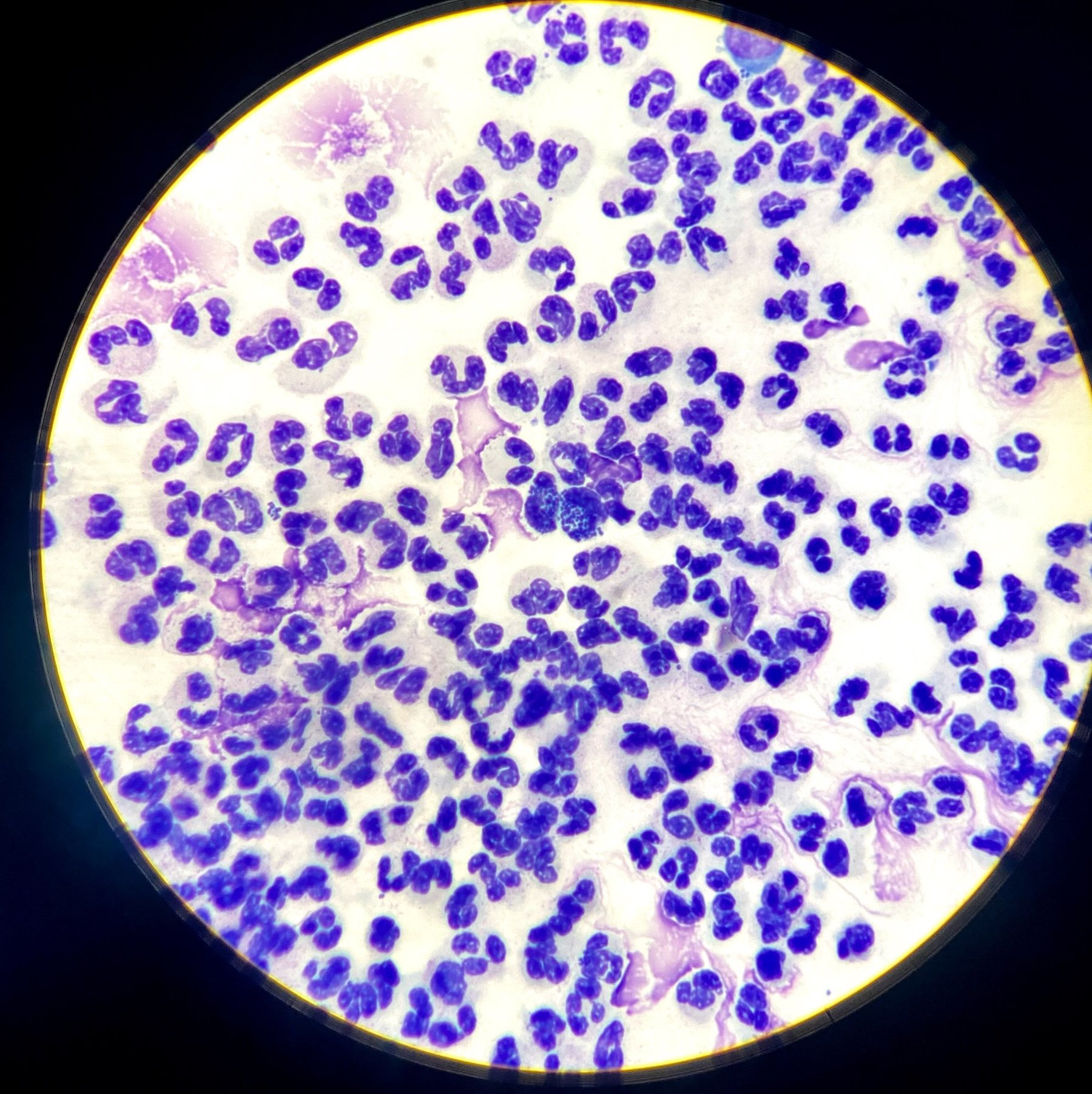
🔬 Urethral smear microscopy (x1000 magnification): Continuous fields of leukocytes are visualized in the preparation. Among them, diplococci are identified, localized both intracellularly (within leukocytes) and extracellularly. The morphology of the diplococci (a pair of adjacent cells, coffee bean shape) and their arrangement are characteristic of Neisseria gonorrhoeae.
This picture requires mandatory confirmation using additional diagnostic methods: ✅ Cultural method (culturing on selective media, e.g., Thayer-Martin medium). ✅ Molecular genetic studies (PCR for Neisseria gonorrhoeae DNA detection).
🔬 The image was kindly provided by laboratory diagnostics physician Aelita Kotova, Moscow.


