

Peripheral blood smear. Normoblasts. So many in the field of view 👀
Photos kindly provided by KDL biologist Yulia Guseva of GBUZ Belorechenskaya CRB.
Morphology of normoblasts in a peripheral blood smear: pronounced normoblastemia, anisocytosis, and polychromasia in a patient's sample from a central district hospital.
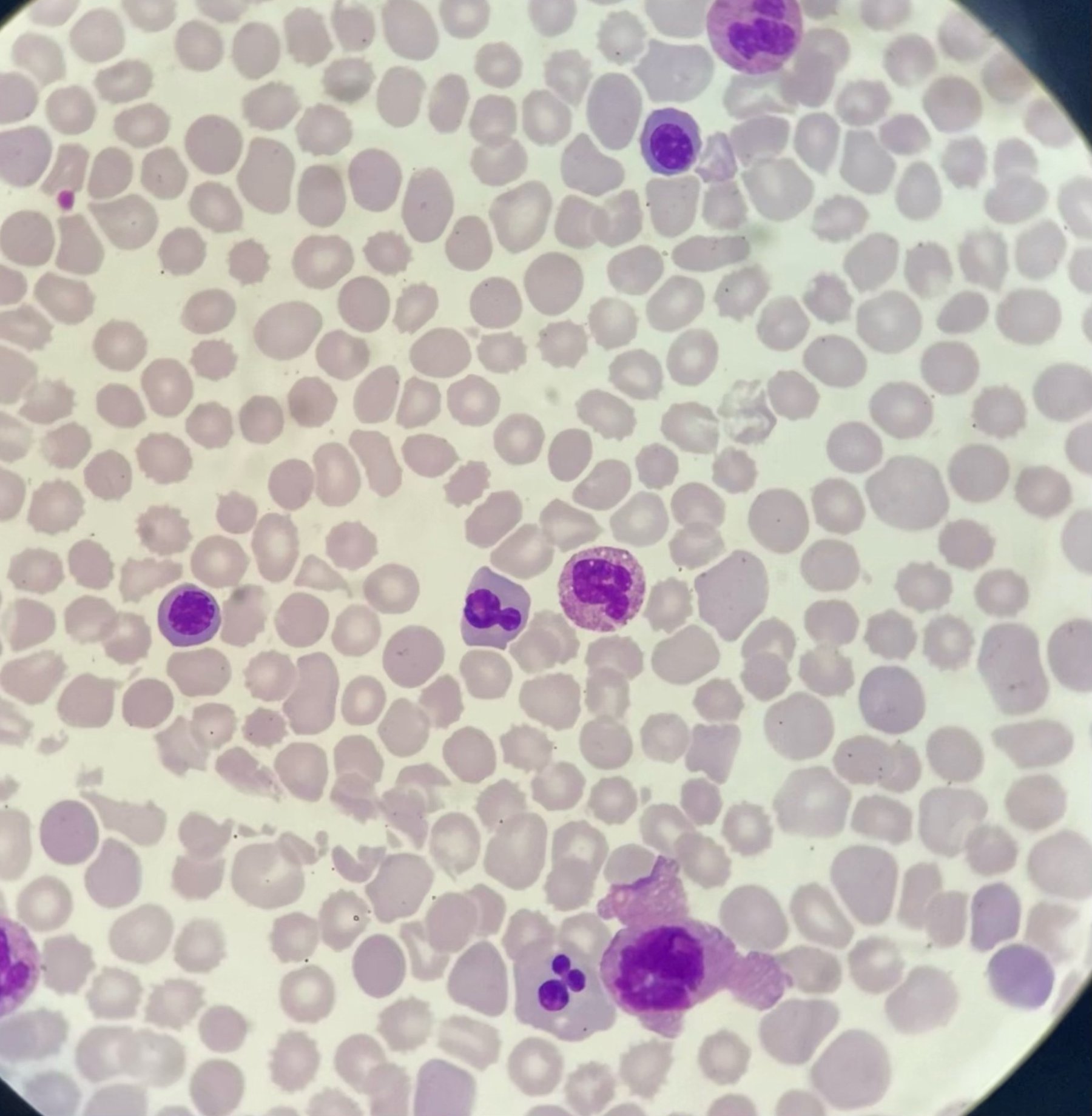
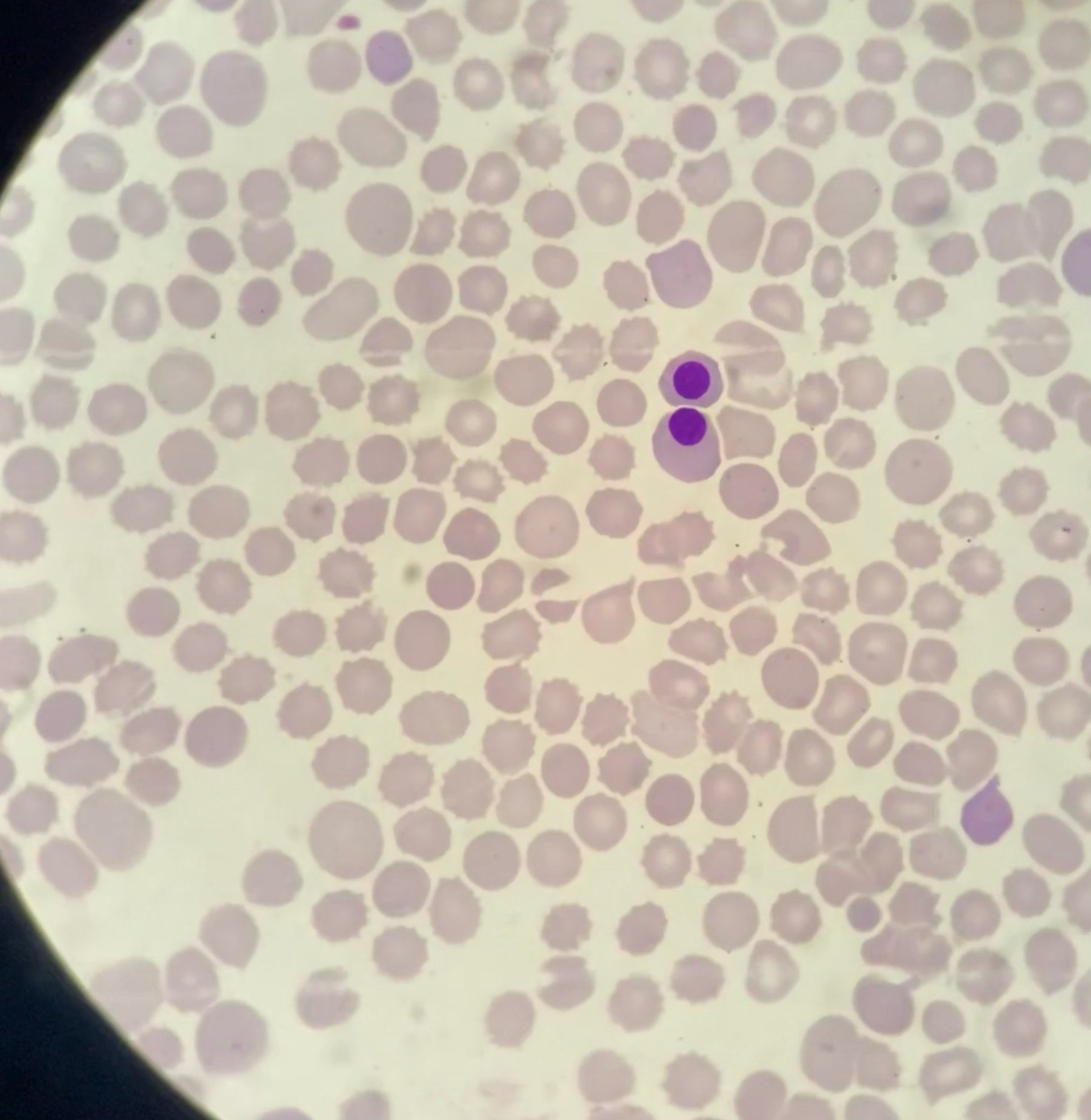
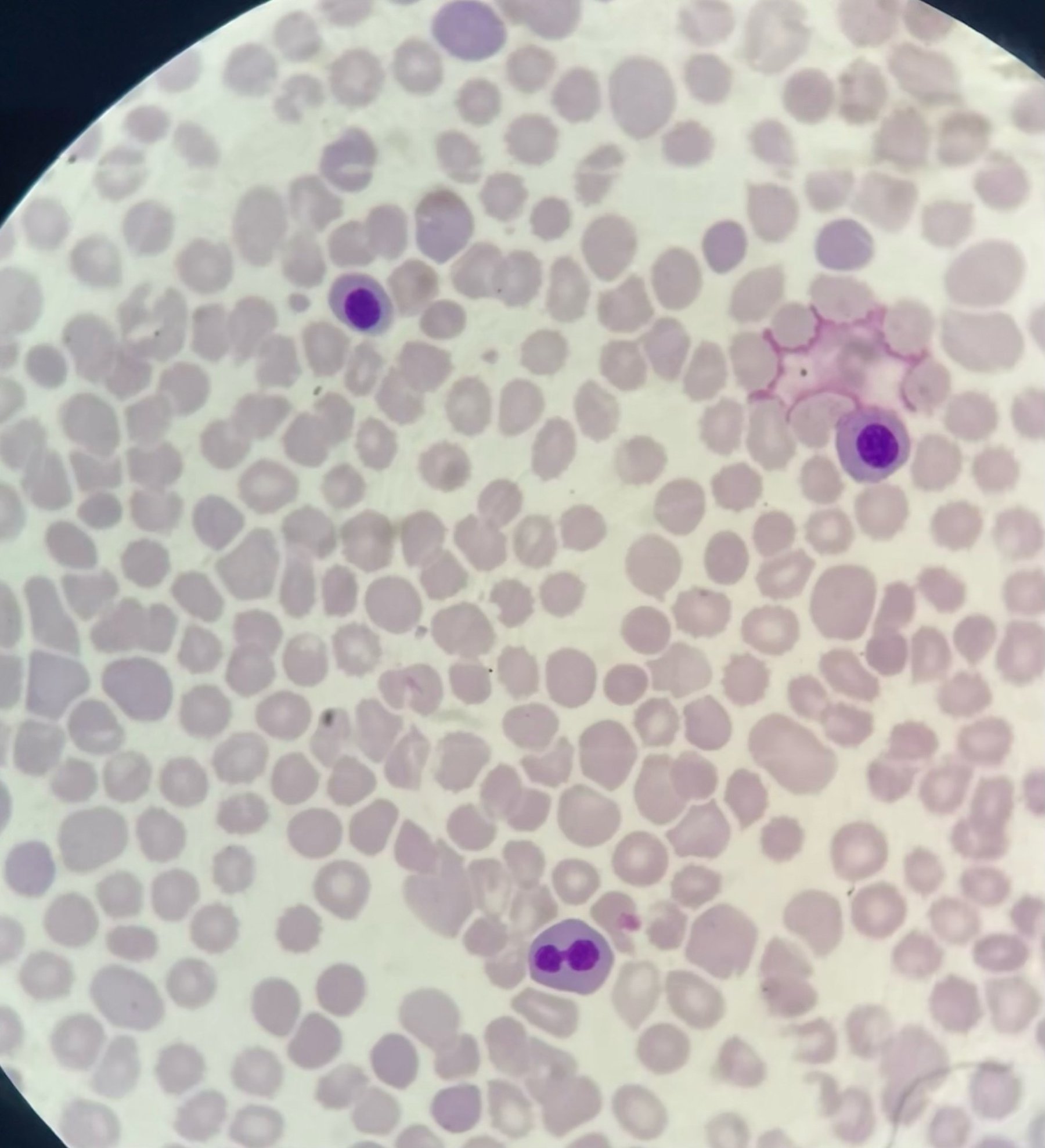
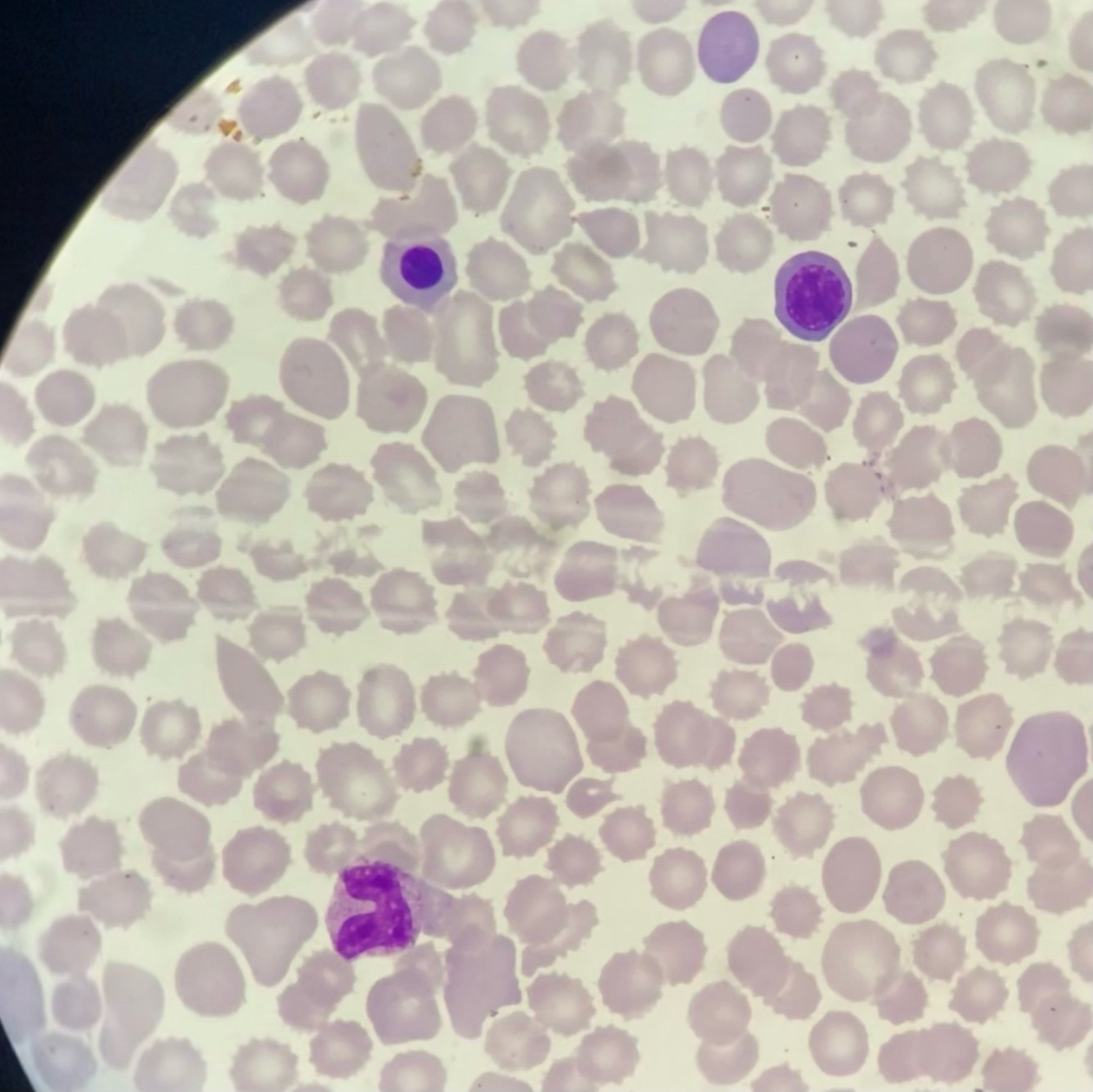
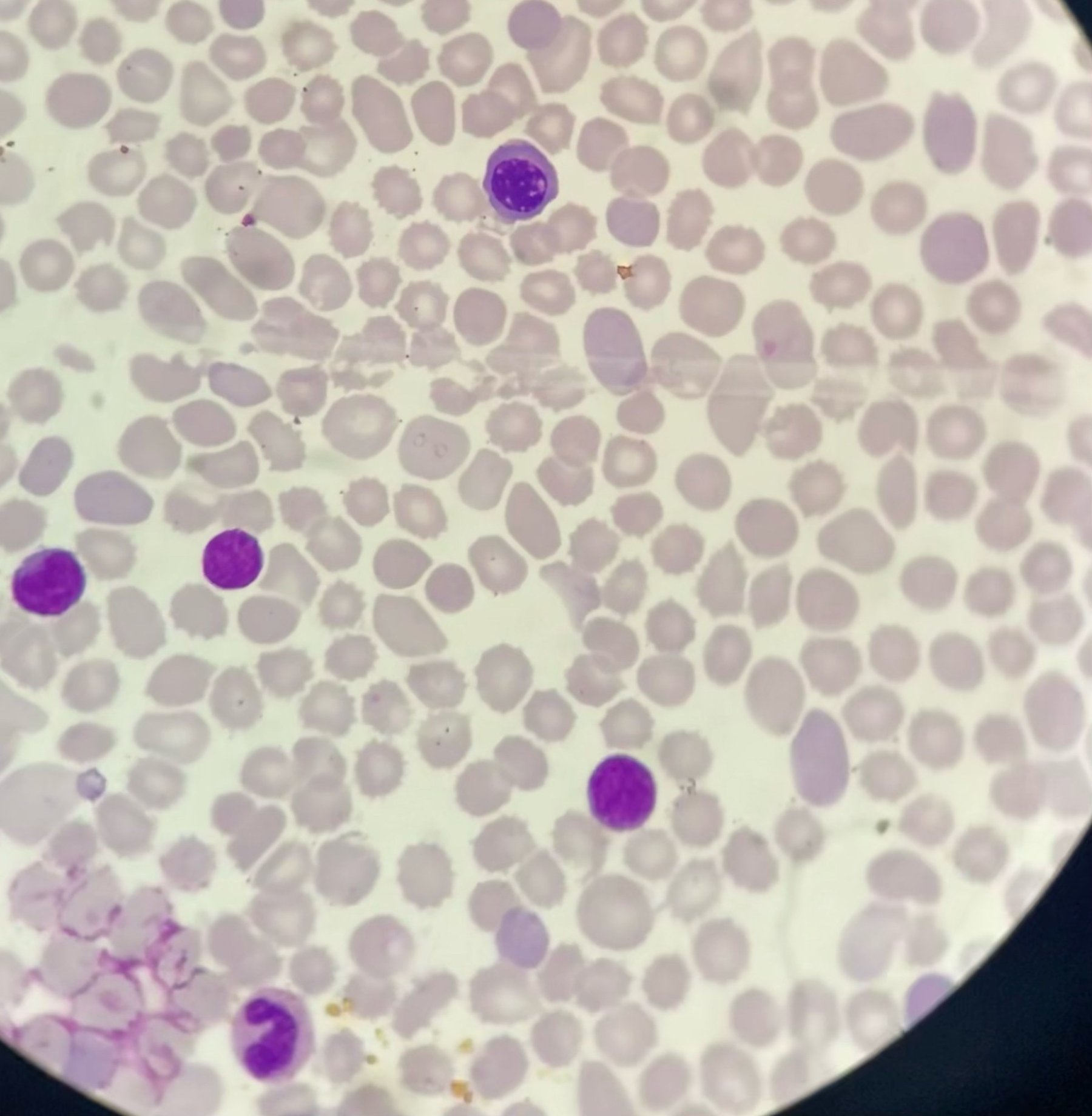
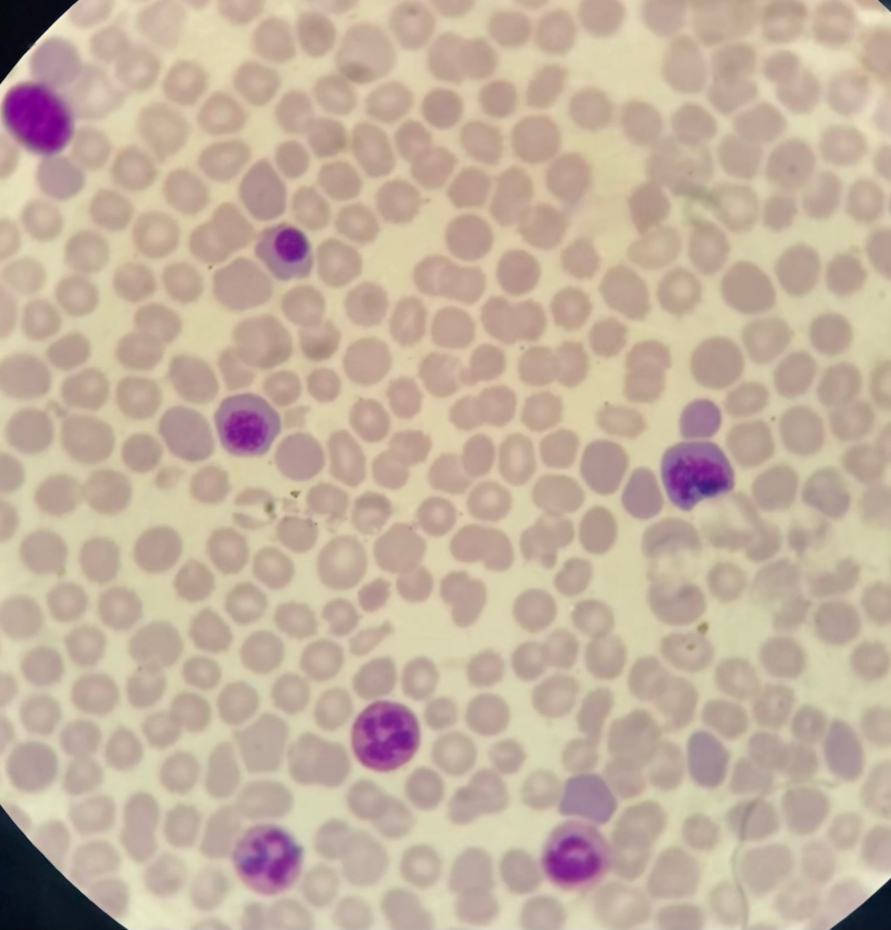
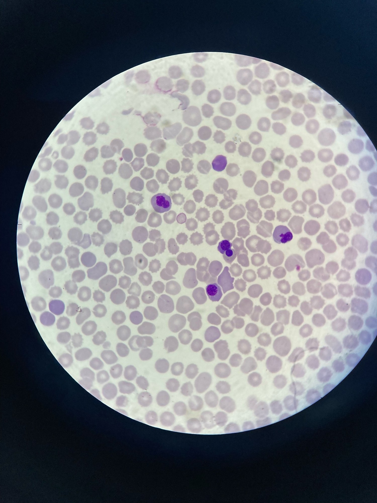
Peripheral blood smear. Normoblasts. So many in the field of view 👀
Photos kindly provided by KDL biologist Yulia Guseva of GBUZ Belorechenskaya CRB.

Clinical case: an internuclear chromatin bridge between normoblasts in the peripheral blood of an ICU patient with multiple organ failure.

A case of severe microcytic anemia with normoblastosis (864/100 WBC): erythrocyte morphology, target cells, Howell-Jolly bodies, and analyzer scattergram.

A clinical case of 91 normoblasts per 100 leukocytes in a peripheral blood smear from a 53-year-old female patient: microscopy and digital morphometry.