

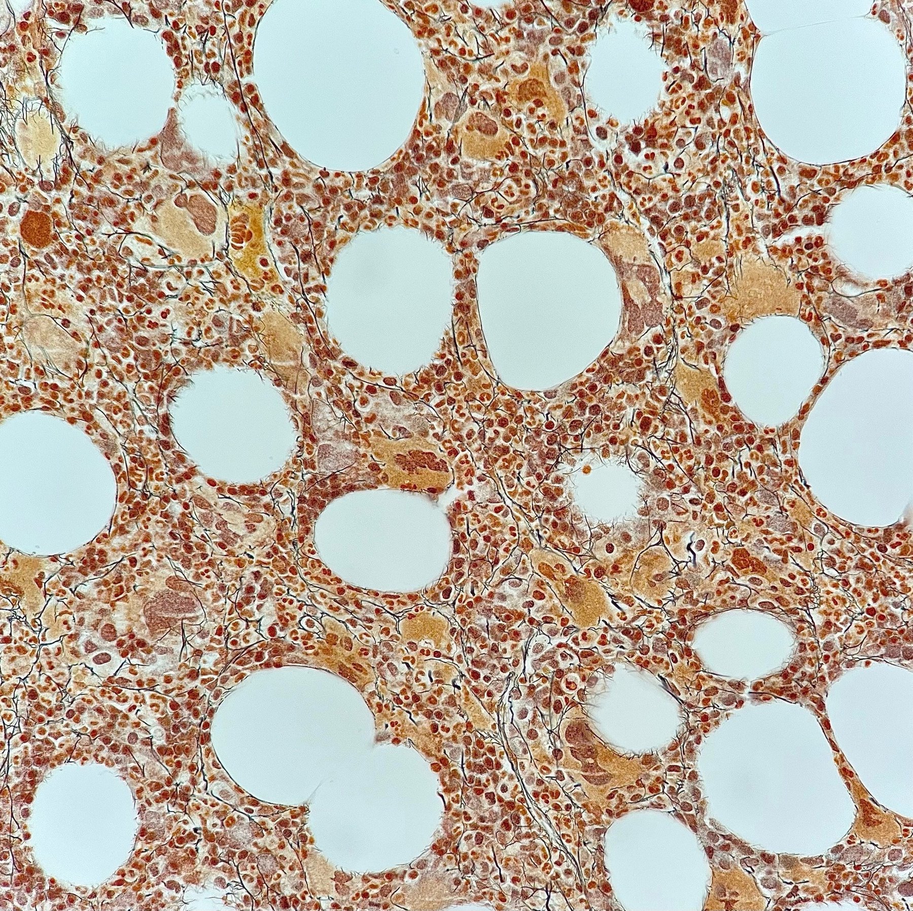
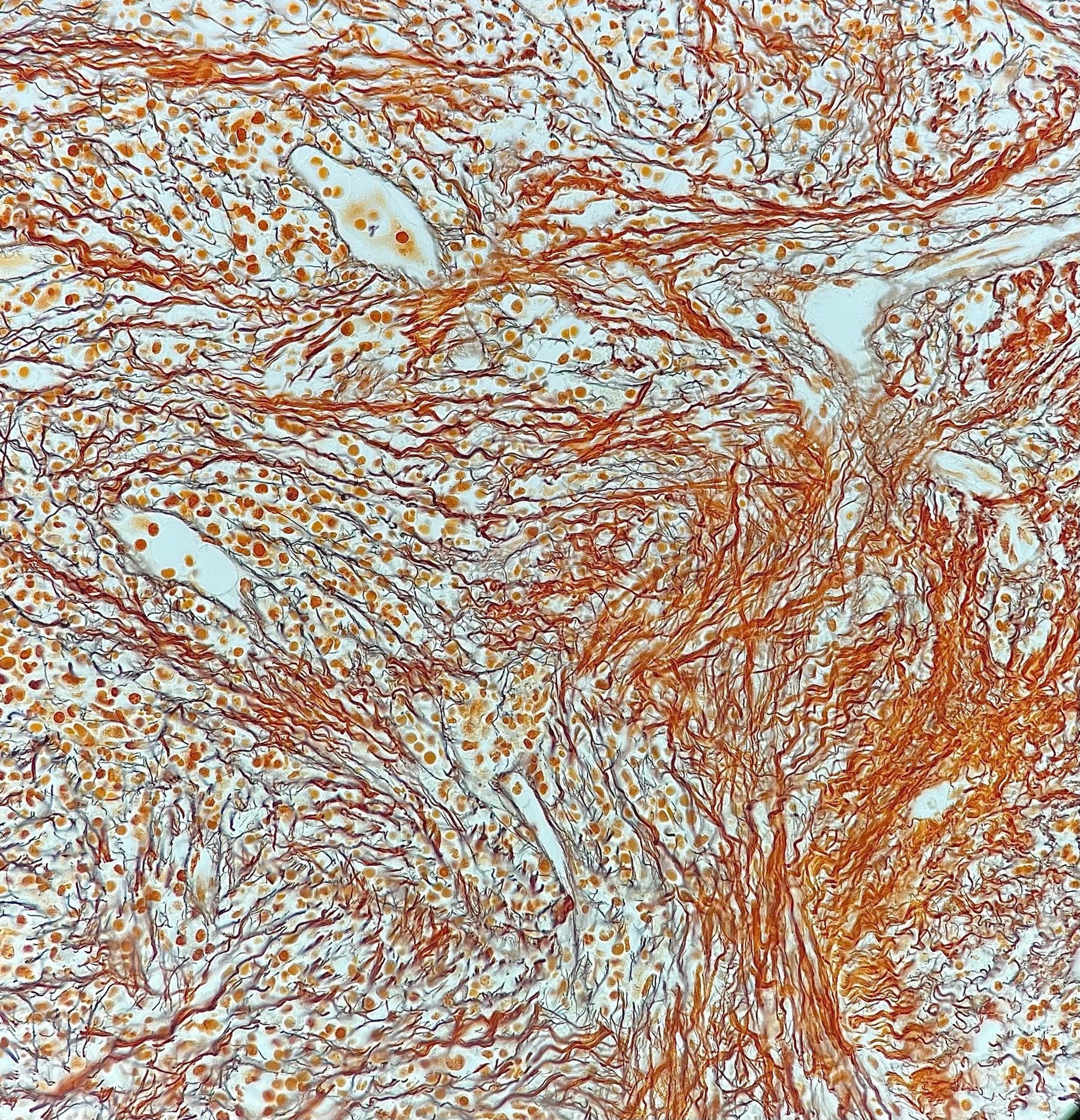
Histochemistry! 🔬Reticulin fibers (Gordon-Sweet stain, silver impregnation).
✅Photo #1. The bone marrow shows areas of MF-1. A loose reticulin network with numerous intersections, especially in perivascular areas. ✅Photo #2: Bone marrow fibrosis, stage: MF-3. Diffuse and dense increase in reticulin with extensive intersections and coarse bundles of thick fibers, corresponding to collagen.
👉The images were kindly provided by Elena Alexandrovna Belyakova, a clinical laboratory diagnostics physician and pathologist from St. Petersburg, Department of Clinical Molecular Morphology, E.E. Eichwald Clinic (Center for Differential Lymphoma Diagnostics), I.I. Mechnikov Northwestern State Medical University.


