

🔬 Rare Diagnosis: How a “Lymphoma Mimic” Confused Doctors?
A 60-year-old man was admitted to the hospital with hematomas and enlarged lymph nodes. Tests showed abnormal “lymphocytes” (86% of all cells), initially leading to a suspicion of lymphoproliferative disease.
🔎 What was found? Upon detailed examination:
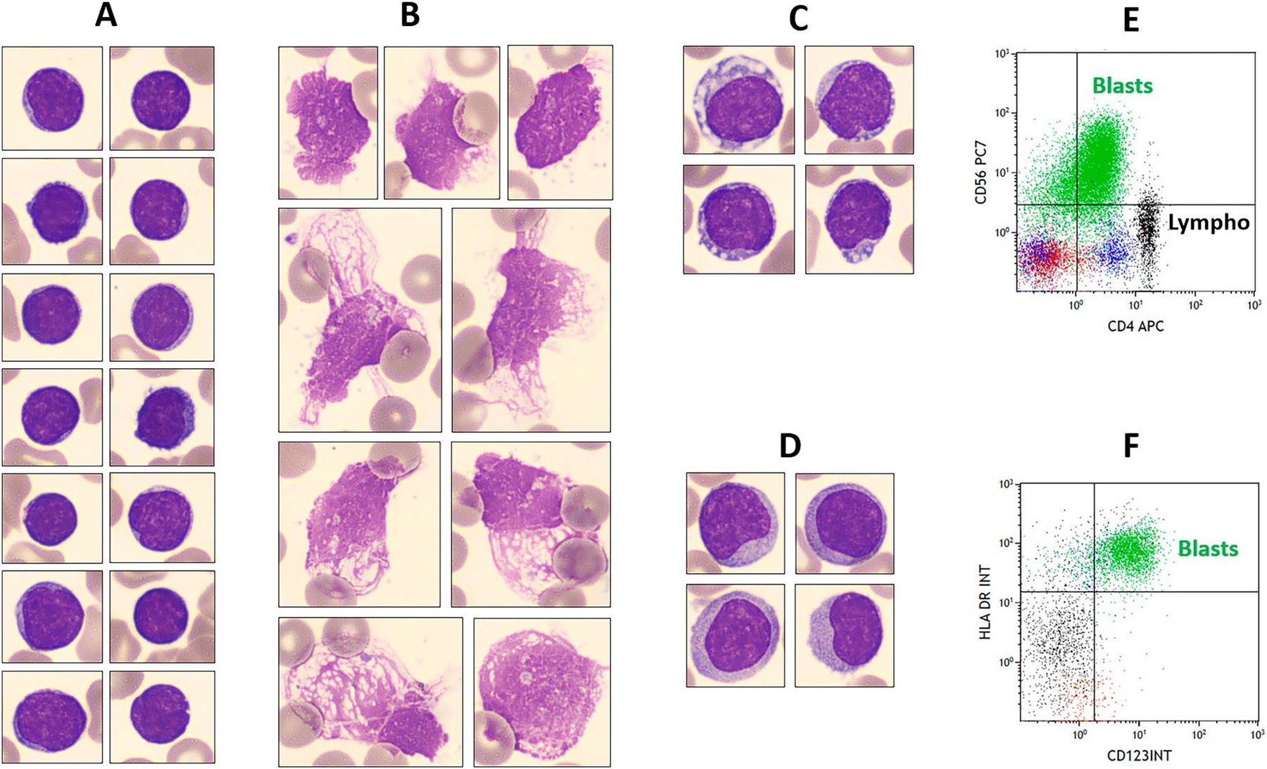
- A blood smear revealed atypical cells — some resembled mature lymphocytes, others had vacuoles or grayish cytoplasm.
- However, flow cytometry (a precise cell analysis method) identified not lymphoma, but a rare blastic plasmacytoid dendritic cell neoplasm (BPDCN).
🧬 Why is this important?
- BPDCN is rare and often “mimics” other diseases.
- In this case, almost all cells looked lymphoid — a rare finding!
- Key to the diagnosis were specific markers (CD56, CD123, HLA-DR) and mutations in TET2, ASXL1, NF1 genes.
💡 Conclusion Even with a “typical” picture, it is important to use additional methods — otherwise, a rare but aggressive tumor can be missed.
Source: Ravel-Chapuis R. et al. American Journal of Hematology. 2025;100:1432–1433.


