

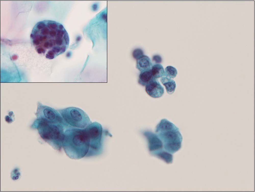
🧪 Why are high-grade cervical lesions missed?
🔬 Diagnostic Cytopathology journal — available by subscription ⭐VK Donut Labformula! Today we uploaded more issues of the journal. Now for 2015 (over 200 publications, over 1000 images!). All available in our LAB-TESTS library.
❗ Why Diagnostic Cytopathology? The journal provides deep, specialized knowledge without “filler”. High-quality images for accurate interpretation. Peer-reviewed articles from world experts.
📚 This is not just reading — it’s your professional growth!
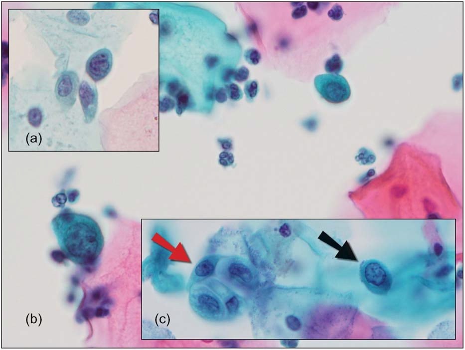
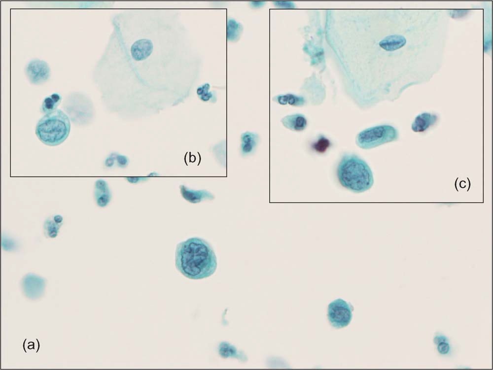
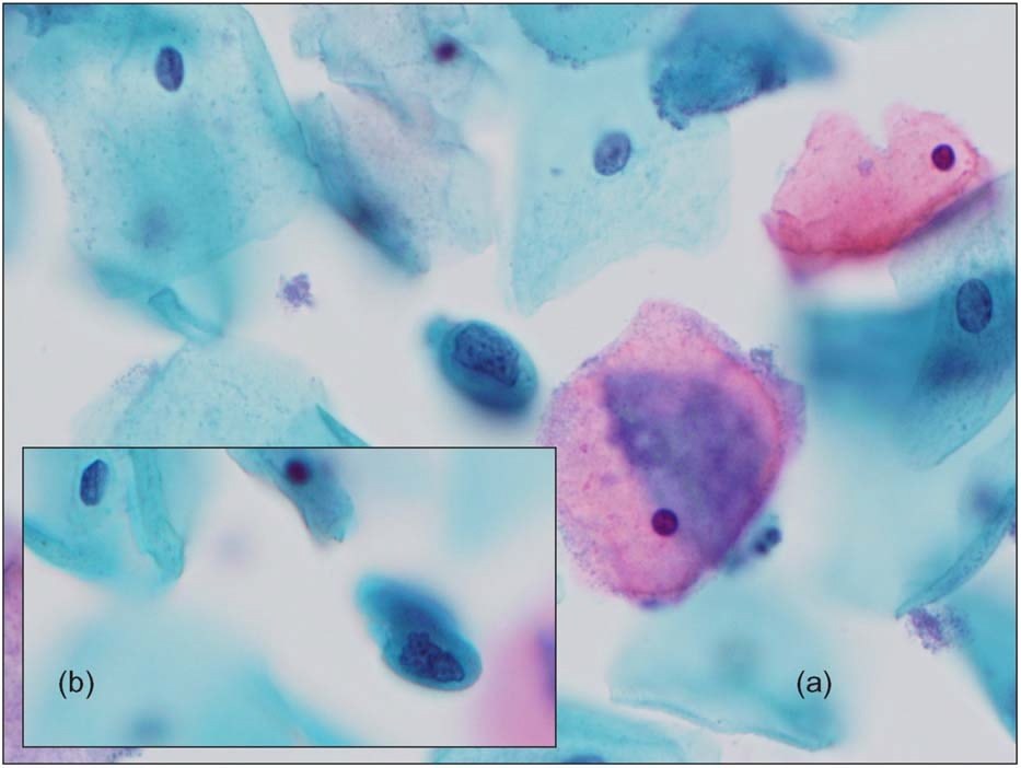
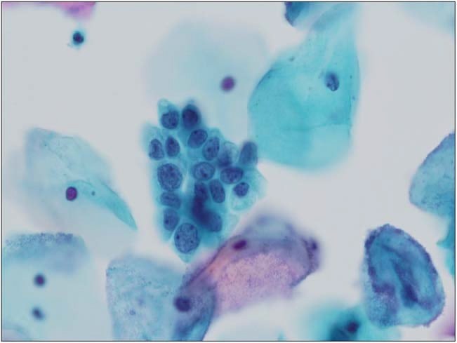
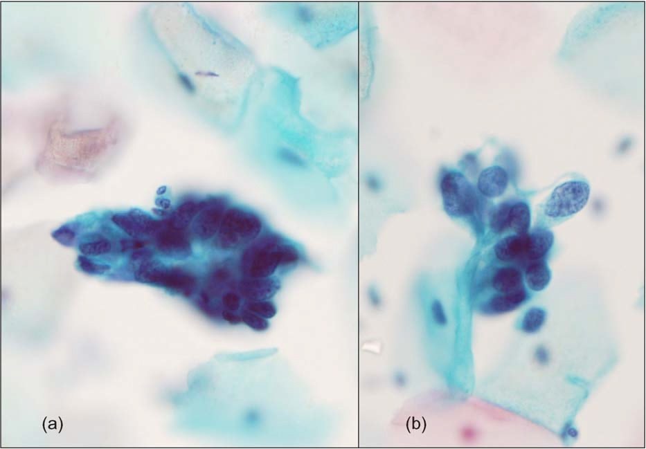
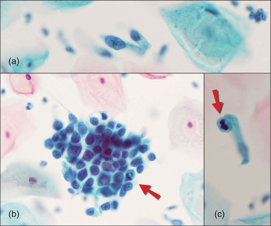
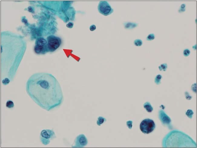
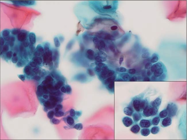
An Australian study analyzed the causes of false-negative results in SurePath® cytological preparations, reviewed using the FocalPoint™ computer system.
🔍 Found:
- HSIL often masquerade as pale, immature metaplastic cells that do not attract attention
- AIS can appear as scattered columnar cells or “open” groups with intercellular spaces
- Endometrial cancer — small 3D groups of cells with bluish nucleoli
📌 Errors occurred even when suspicious cells were present in the field of view — they were not recognized or underestimated 📸 The work emphasizes the need to focus and scan the entire field, not just the center
📈 Conclusion: morphology in SurePath requires a new recognition strategy — non-obvious signs can be key to early diagnosis!
🔗 Source: Bowditch RC et al. Morphologic Analysis of False Negative SurePath® Slides Using FocalPoint™ GS Technology. Diagnostic Cytopathology. 2015;43(11):870–878.


