

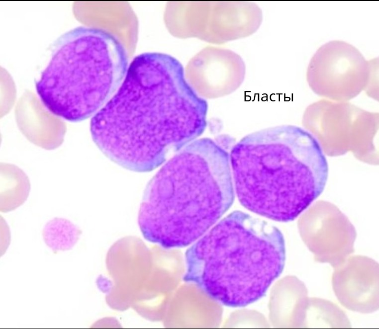
The correct answer to the closed club of laboratory hematologists’ challenge. D) 3 errors
🧾 Explanation: The resident made three serious errors:
There is no leukopenia — leukopenia cannot be diagnosed without a complete blood count, especially from a single field of view of a smear.
There are no reactive lymphocytes — there are no lymphocytes in the smear, let alone reactive ones. All cells are blasts, which is characteristic of hemoblastoses, not viral infections.
This is not “viral blood” — neither the cytological picture nor the cellular composition of the smear shows signs of viral infection. The conclusion about a “patient with a virus” is fundamentally erroneous.
📌 In such a situation, the correct conclusion is a suspicion of acute leukemia and urgent further investigation.
🏆 Congratulations to the winners! They receive a private invitation to the club of selected specialists.
- Olga Borodich - membership in the club confirmed
- Anastasia Belsheva - invitation sent to DM
- Tatyana Kudryashova - invitation sent to DM
- Anastasia Ivanova - invitation sent to DM


