

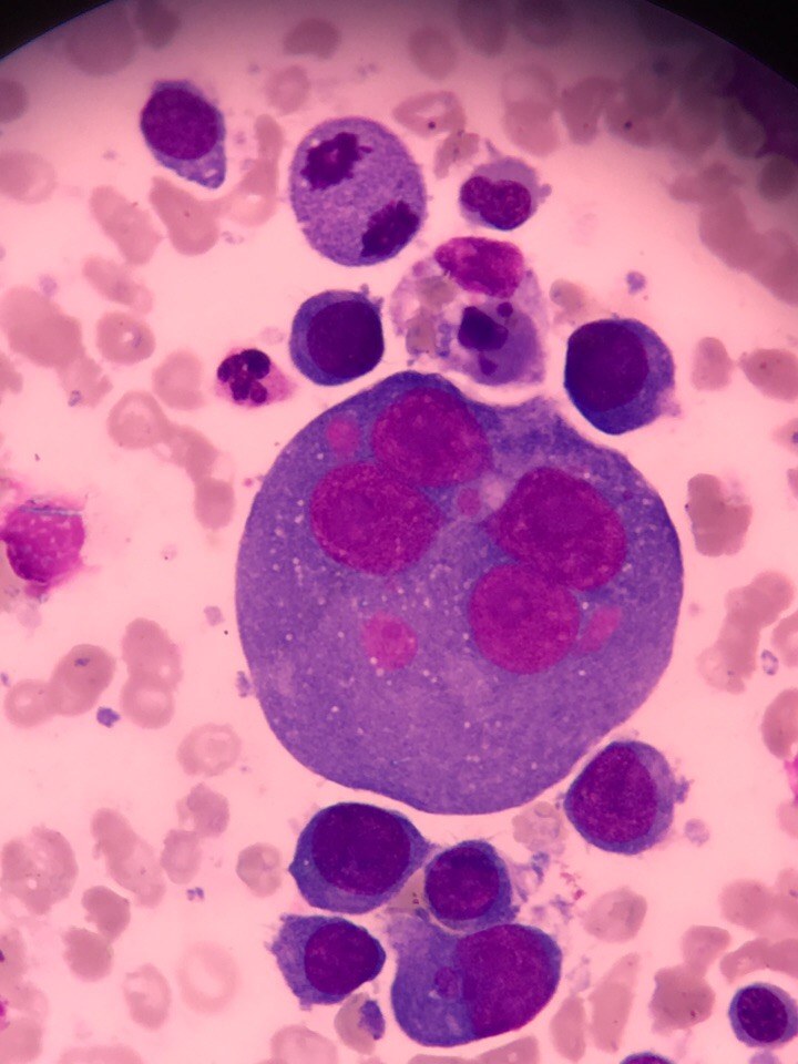
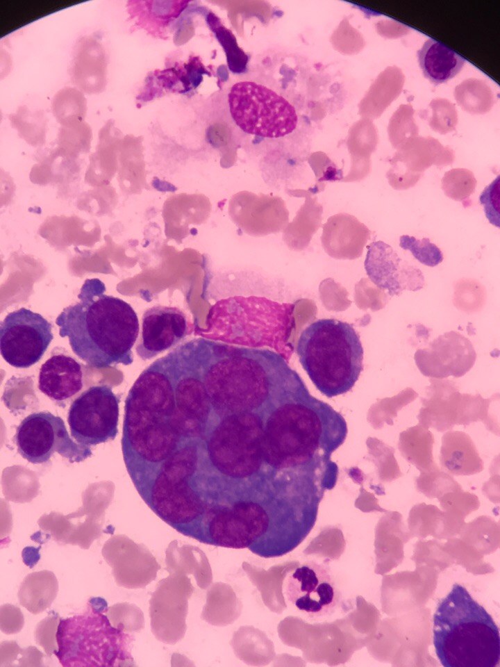
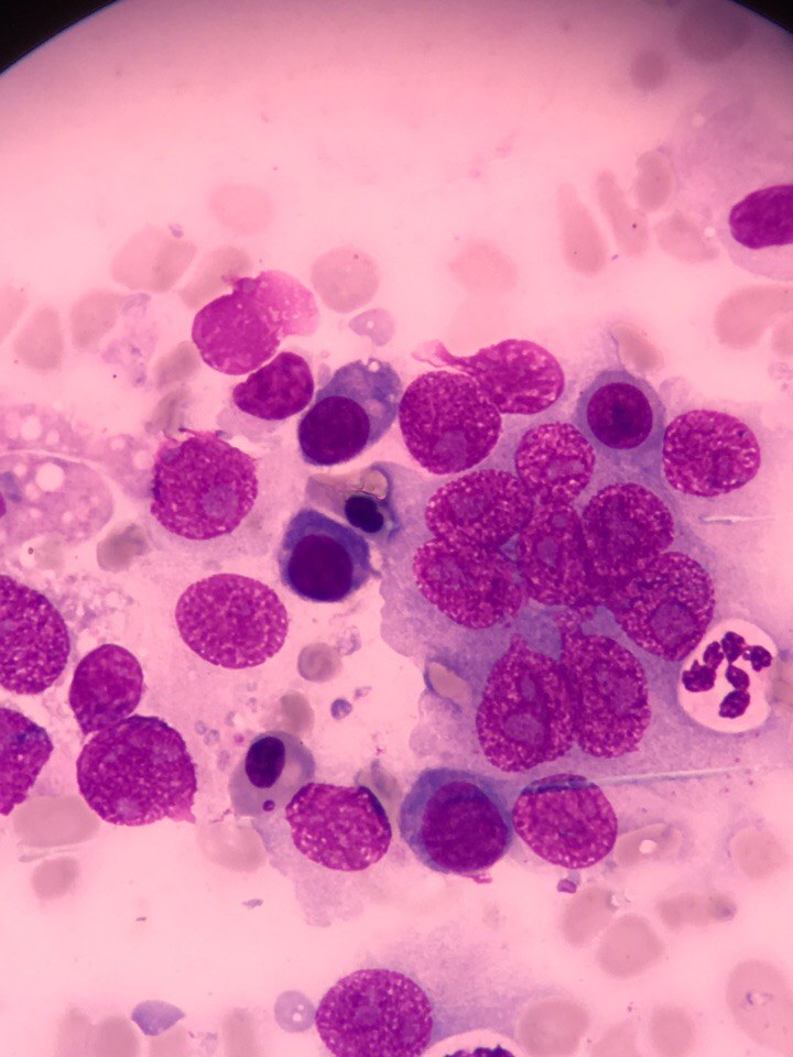
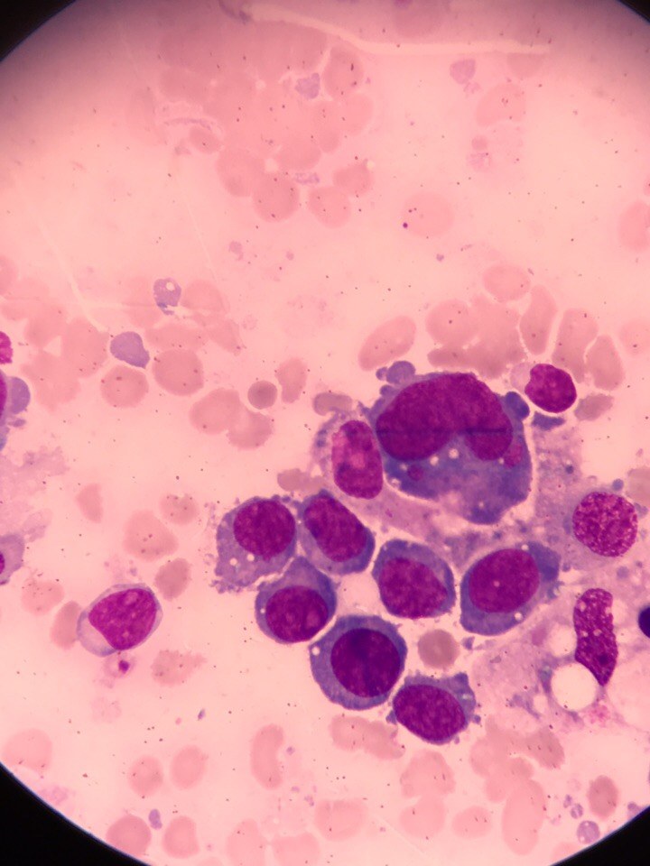
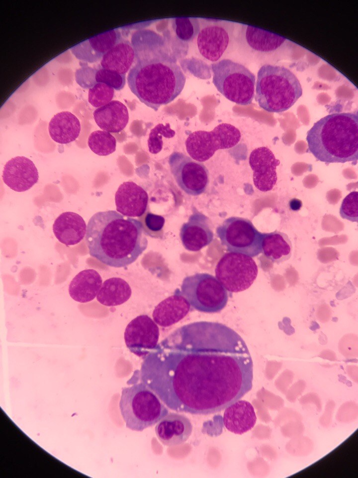
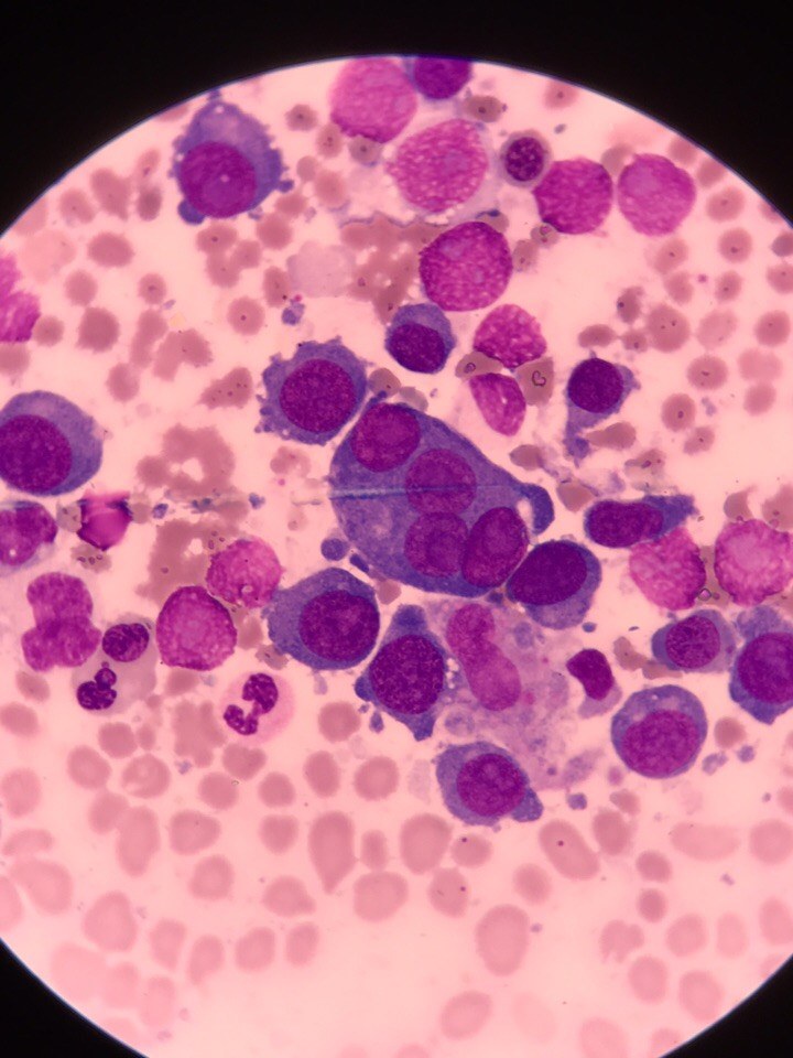
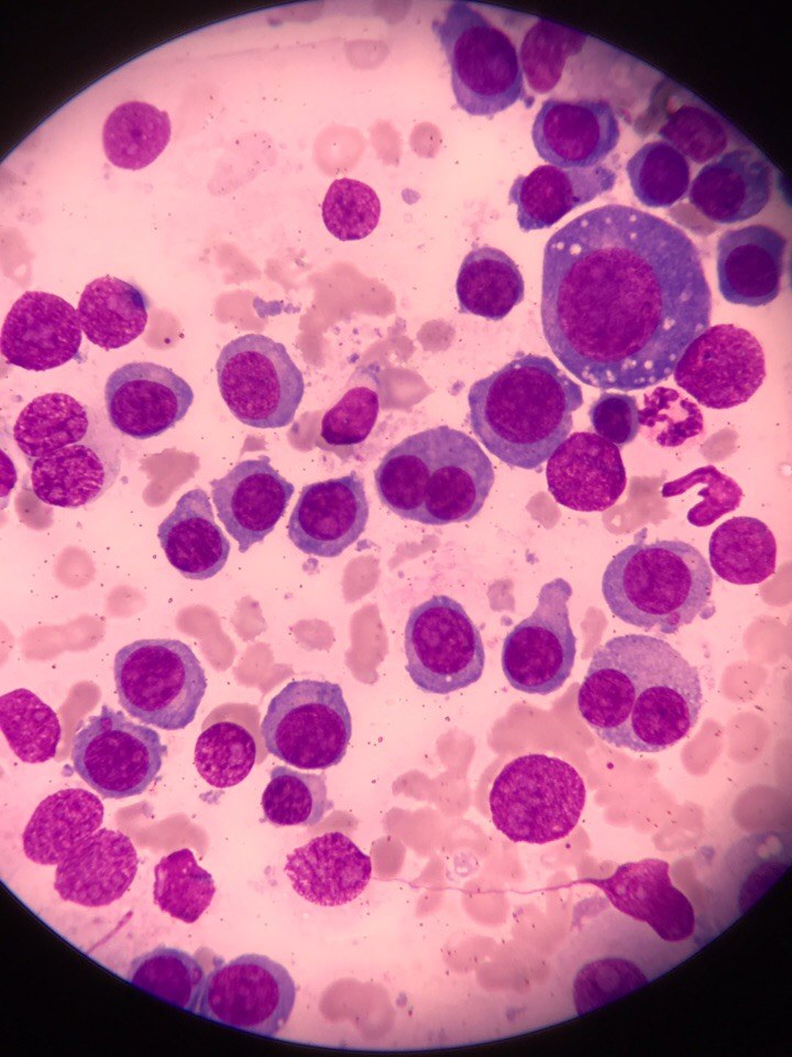
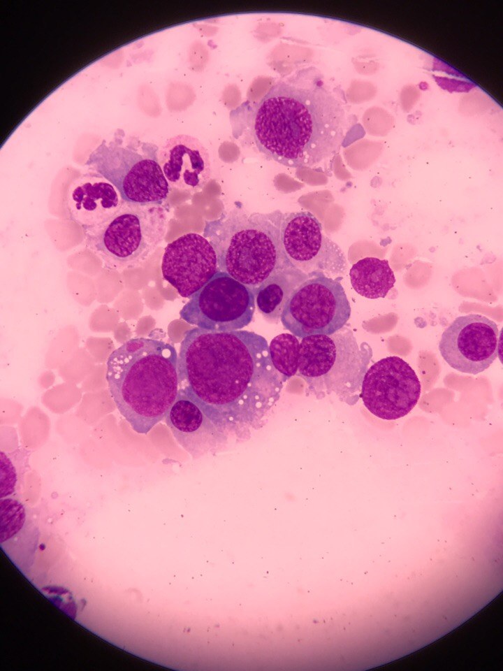
In a 65-year-old woman admitted to the hematology department. CBC: leukocytes-15.40x10⁹/L, platelets-46x109/L, HGB-86 g/L, erythrocytes-2.85x10^12/L. The hematological analyzer did not count the differential. Peripheral blood smear microscopy: blasts-12, myelocytes-5, metamyelocytes-2, band neutrophils-2, segmented neutrophils-53, monocytes-23, lymphocytes-3. Normoblasts per 100 leukocytes-45.
In the bone marrow aspirate: blasts-60%, erythroid cells 23%, of which 5% are megaloblasts.
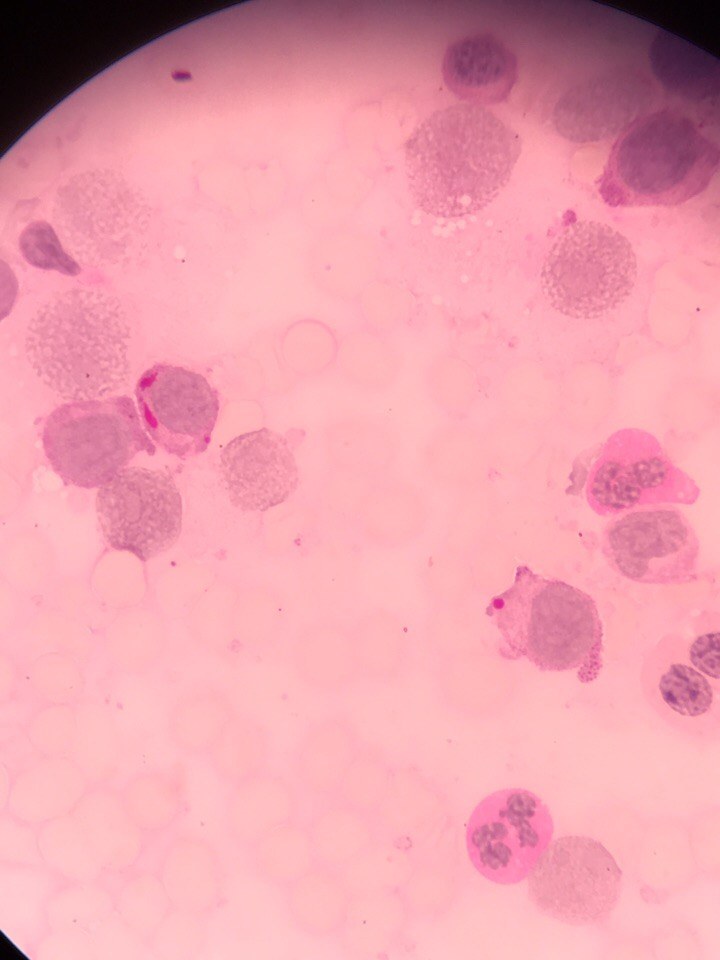
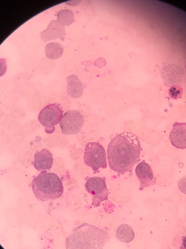
Upon examination of the slide, naked nuclei were about 24%, in some areas more. Megakaryocytes were found in sufficient numbers. Blastic cells of various sizes (from small to giant), mononucleated and multinucleated (gigantoblasts) with a high nuclear-cytoplasmic ratio. Proliferation of erythroid cells with pronounced signs of dyserythropoiesis (multinucleated, giant normoblasts, cytoplasmic bridges, megaloblastoid nuclei, karyorrhexis, Jolly bodies) was noted. Cytochemistry: lipids not detected in blast cells, glycogen: 4%-diffuse, 71%-fine granular, 5%-coarse granular on a diffuse background. Some normoblasts contain glycogen.
According to IFT: M6


