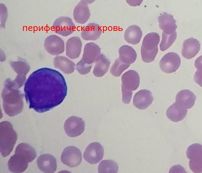
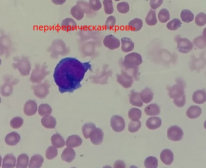
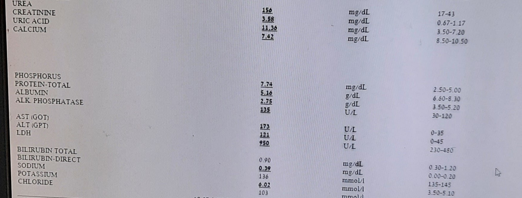
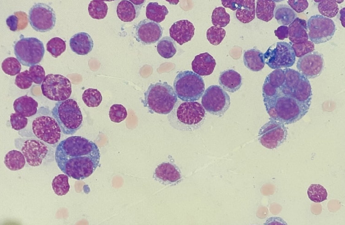
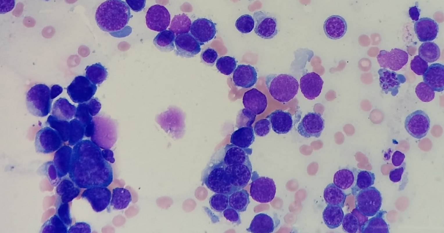
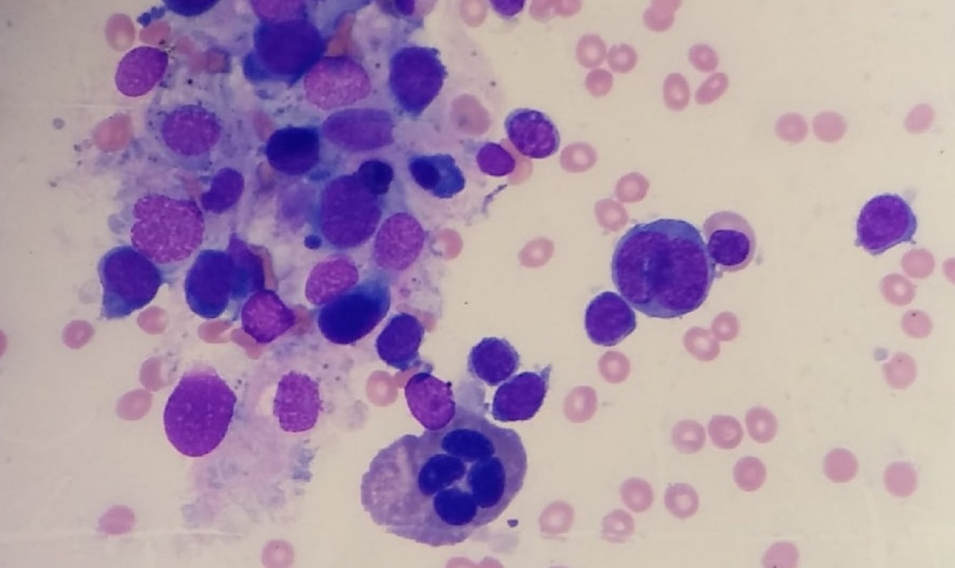
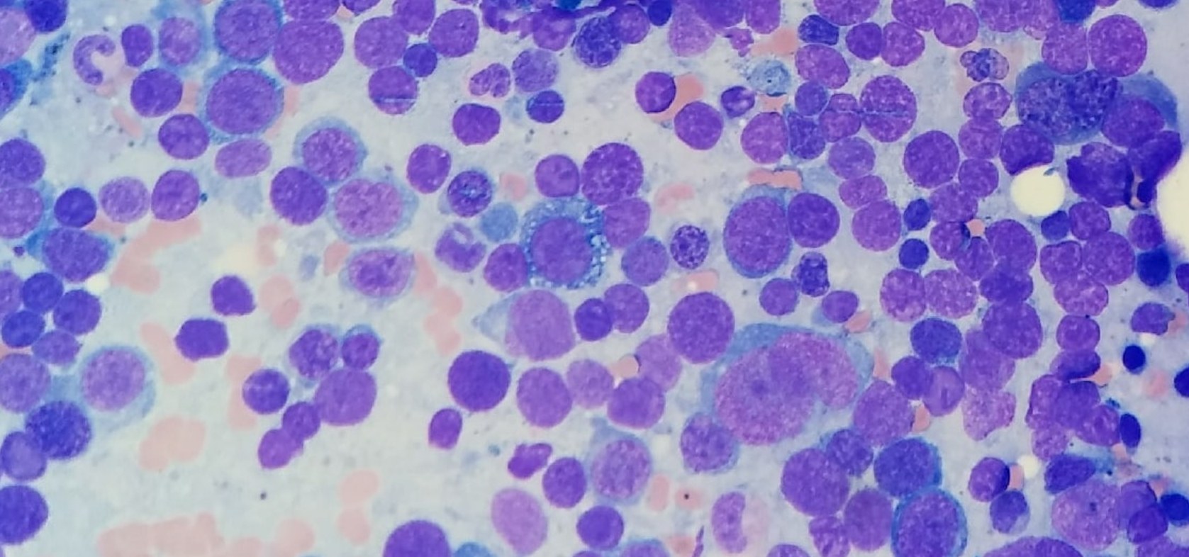
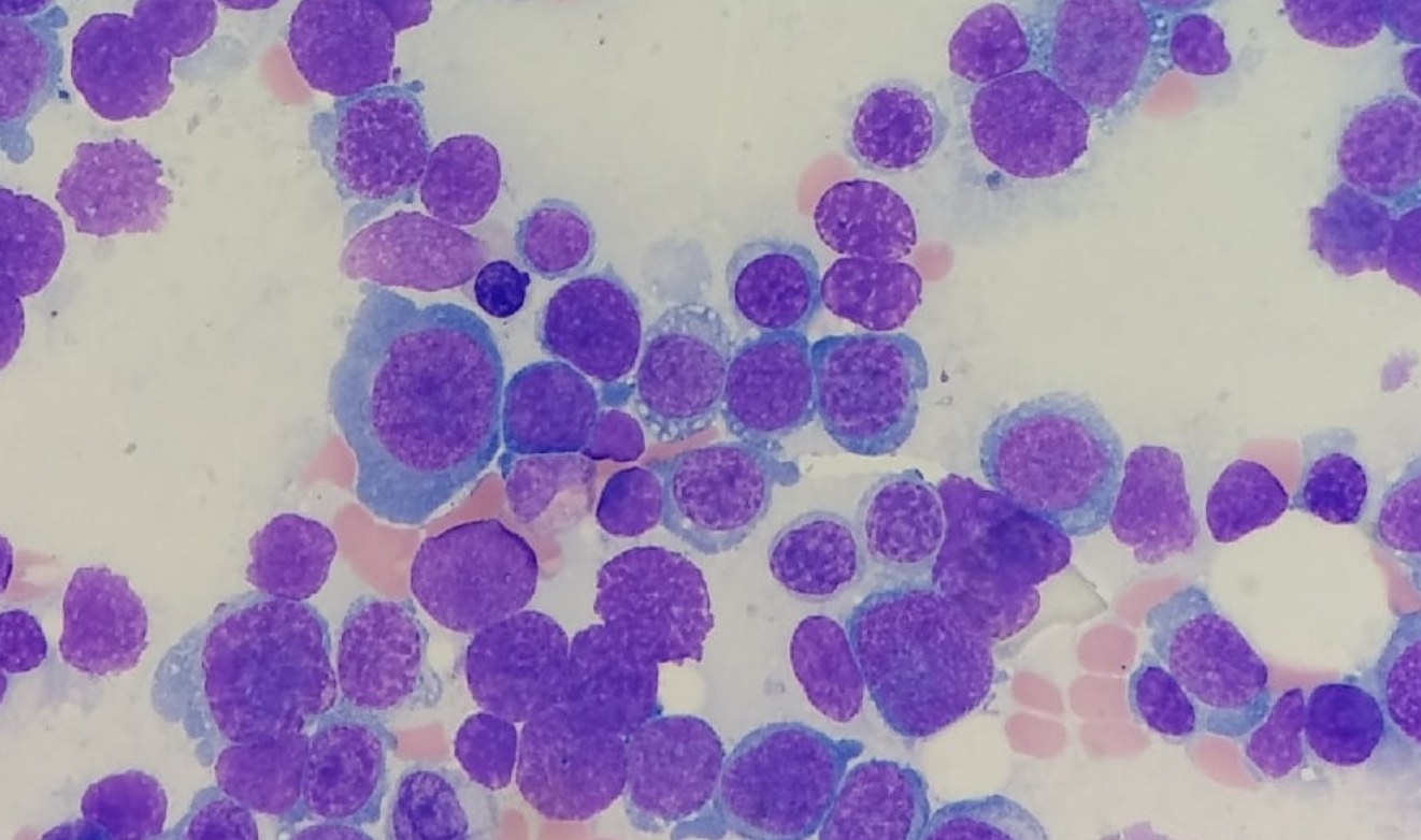
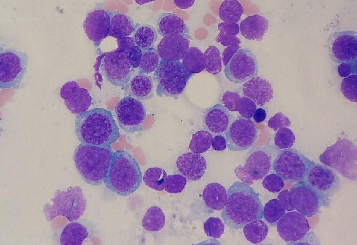
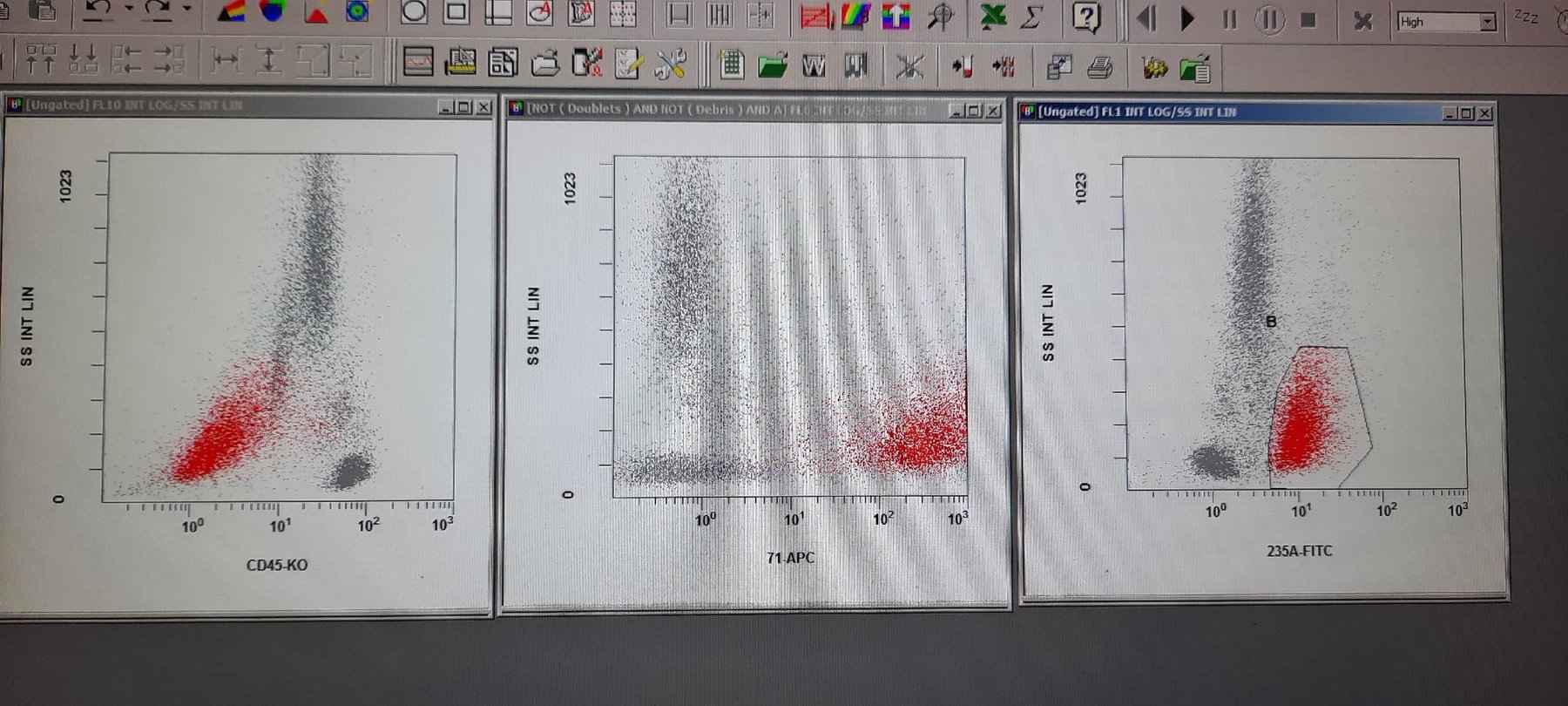
👉Clinical case - acute erythroid leukemia. ✅A 92-year-old male was admitted to the emergency department with retrosternal pain and dyspnea, suspected acute heart failure - troponin 198 ng/L (N=0-14). Hepatosplenomegaly was also detected. ✅Pancytopenia in peripheral blood: Leukocytes (WBC) 3.01×10⁹/L, Hemoglobin (Hb) 59 g/L (normochromic normocytic), Platelets (PLT) 9×10⁹/L. ✅Single blast-like cells in the peripheral blood smear (see photo). ✅Bone marrow aspirate shows marked proliferation of immature erythroid elements with some dysplasia (multinuclearity, cytoplasmic vacuolization). ✅Immunophenotyping revealed a CD45 negative population, positive for erythroid markers CD71, CD235a, CD36, and negative for CD34, CD117, MPO, CD38, CD41, CD61, CD64, CD33, HLA-DR. ✅Histology conclusion: acute erythroid leukemia.
Acute Erythroid Leukemia: Morphology and Immunophenotype
A clinical case of acute erythroid leukemia: pancytopenia, blast cells in the smear, erythroid dysplasia, and flow cytometry (CD71+, CD235a+).