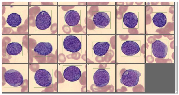
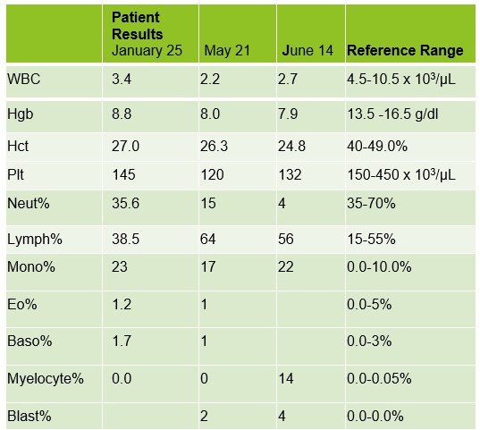
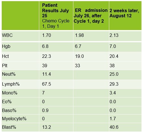
🔬IN THE PHOTO. Peripheral blood smear. Cells classified as blasts in CellaVision from a 75-year-old man with leukopenia. Also in the tables are the dynamics of CBC changes over one year. The blast phenotype was CD13+, CD33+, CD117+, and HLA-DR+. Blasts were negative for CD34. The patient’s diagnosis: acute myeloid leukemia (AML). The drug prescribed was Vidaza (azacitidine). 👉🏻Also of interest is the presence of a group of large granular lymphocyte (LGL) T-cells in the peripheral blood. This is an increased population of CD57+, CD3+ T-cells. LGL clones have been described in AML, and a hallmark of this association is the cytopenia observed in this patient. 👉🏻Note the sharp increase in blast count over a 2-week period from July to August. The disease progressed rapidly. 👉🏻Elderly patients like this generally tolerate traditional aggressive chemotherapy poorly due to other health issues. This patient was likely not a good candidate for strong chemotherapy due to his age and medical history. Source: labmedicineblog
Acute Myeloid Leukemia: Blast Morphology in Blood Smear
A clinical case of acute myeloid leukemia: blast morphology in blood smear, immunophenotype, co-occurring LGL clone, and one-year hemogram dynamics.