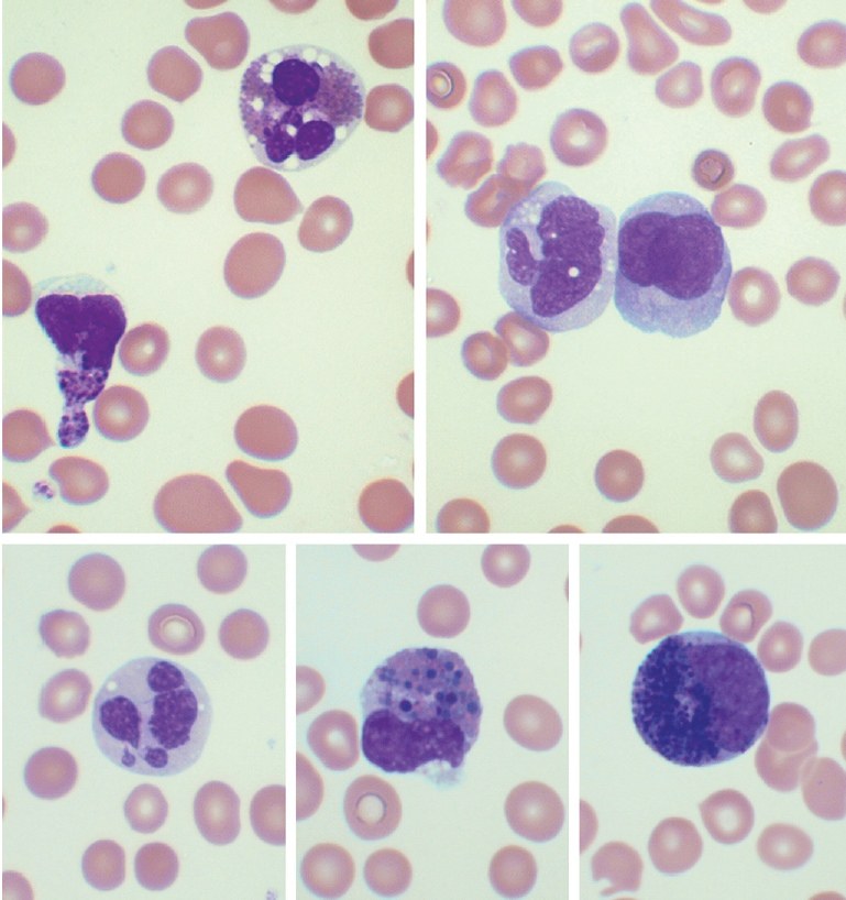
❗ACUTE MYELOID LEUKEMIA (AML) with inv(16) (p13.1q22) ✅These images are from the blood smear of a 48-year-old Somali woman. Blood tests showed: white blood cells 74.9 × 10⁹/L, hemoglobin 102 g/L, and platelets 46 × 109/L. ✅Acute myeloid leukemia (AML) with inv(16) (p13.1q22) can usually be suspected based on characteristic bone marrow morphology. However, careful examination of the blood smear can also reveal distinctive features. The findings are consistent with acute myelomonocytic leukemia with eosinophilia. Although most eosinophils show only minor dysplastic changes, such as cytoplasmic vacuolation and granule reduction, there may also be some eosinophils and precursors with purple pro-eosinophilic granules, sometimes large. In addition, neutrophils are dysplastic. ✅Cytological abnormalities were typical for this condition. Most eosinophils showed signs of mild dysplasia, such as vacuolation, seen in the top cell in the upper left image. A smaller proportion of cells were more abnormal, being very small or showing an absence of nuclear lobulation or the presence of pro-eosinophilic granules (as seen in the bottom cell in the upper left image). In addition, there were immature monocytes and promonocytes (both shown in the upper right image), monoblasts, and myeloblasts. All neutrophils were very hypogranular with abnormally shaped nuclei (bottom left), often with rounded nuclear lobes, although classic Pelger-Huët forms were not observed. However, the most distinctive features were the small number of circulating eosinophil precursors. Some of these had both mature eosinophilic granules and abnormally large dark purple pro-eosinophilic granules (bottom center); this cell is virtually pathognomonic for this cytogenetic abnormality. Other eosinophil precursors had only pro-eosinophilic granules (bottom right). ✅Recognizing the typical features of AML with inv(16)(p13.1q22) or t(16;16)(p13.1;q22) is important to ensure appropriate investigations are performed. Usually, the relevant abnormalities are detected by cytogenetic analysis, but otherwise, fluorescence in situ hybridization (FISH) is required. Source: Bain Barbara J. - Hematology - 101 Morphology Updates, 2023
Acute Myeloid Leukemia with inv(16): Blood Smear Signs
Morphological features of peripheral blood cells in AML with chromosome 16 inversion: eosinophil dysplasia, promonocytes, and neutrophil hypogranulation.