

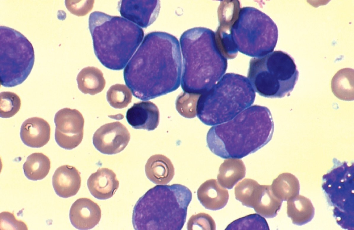
A 12-year-old boy suddenly developed fatigue, which progressively worsened. At birth, he was diagnosed with Down syndrome and intellectual disability. Physical examination revealed physical abnormalities associated with Down syndrome, pale mucous membranes, and mild splenomegaly. There was no lymphadenopathy or hepatomegaly. The doctor ordered a complete blood count and urinalysis. ✅ Laboratory data. Red blood cells and hemoglobin were sharply reduced. The total white blood cell count was 255×10^9/L. In the blood smear: blast cells 82%, promyelocytes 2%, myelocytes 2%, metamyelocytes 1%, segmented neutrophils 7%, lymphocytes 6%. Platelet levels were also significantly reduced. Auer rods were noted in many blasts. Urinalysis was normal. Subsequent cytochemical staining showed that some blasts were positive for Sudan black B. Negative results were obtained with alpha-naphthyl acetate esterase (monocytic esterase), PAS, and acid phosphatase.
What is the most likely diagnosis?


