

A case of diagnosing 500,000 leukocytosis!
After processing a blood sample from an outpatient clinic (male, 71 years old) and obtaining this result, doubts arose about its reliability: Leuk. 503×10⁹/L (the analyzer’s cell distribution across all possible channels only added to the uncertainty), Hgb 85 g/L, Platelets 196×10⁹/L. We repeated the analysis, while a smear was prepared and the patient’s history was reviewed. The result was repeated, and in June 2022, the patient was diagnosed with Diffuse Large B-cell Lymphoma of the soft tissues of the back. He underwent chemotherapy. At present (end of July 2023), we have no clinical data.
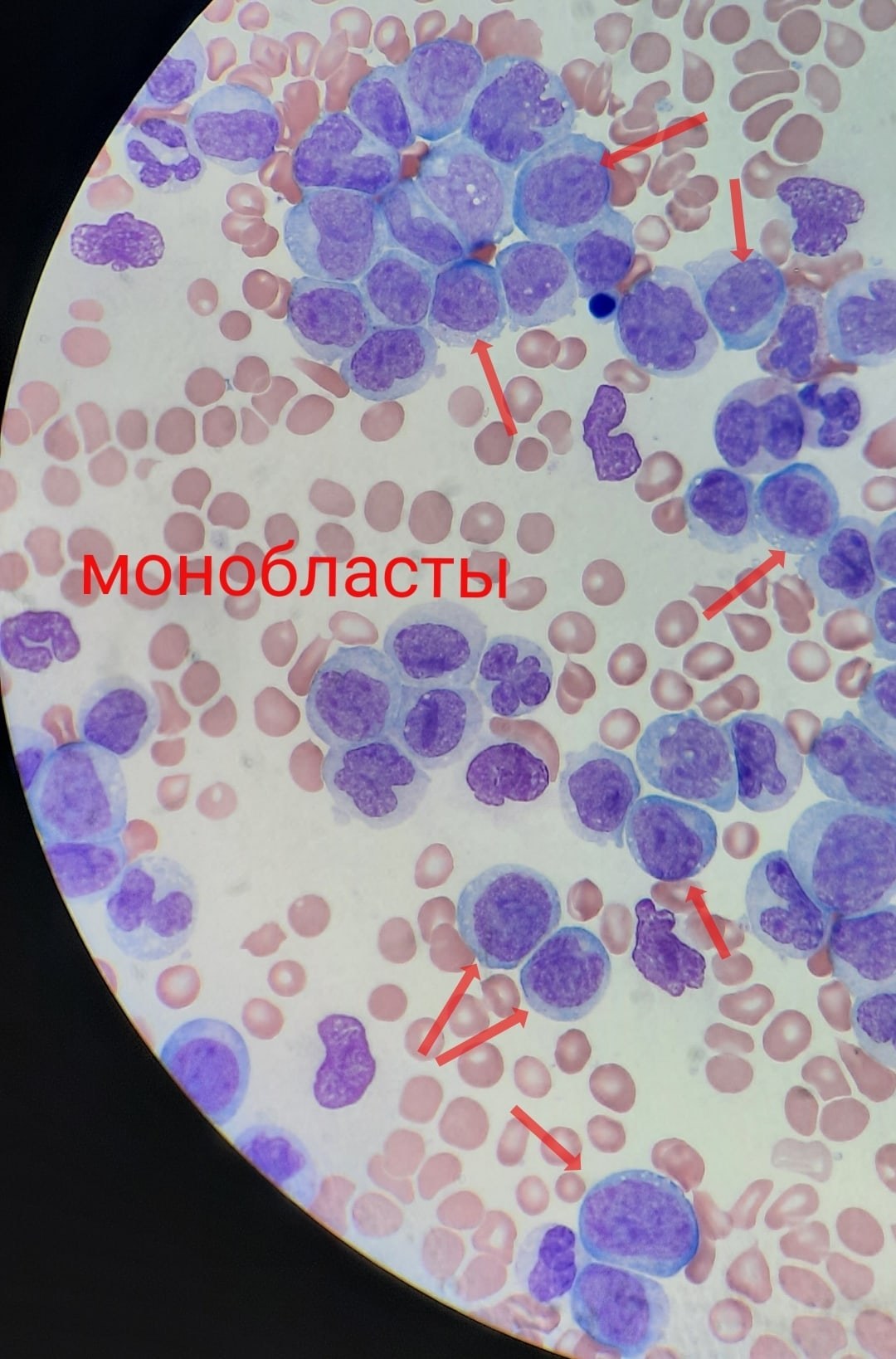
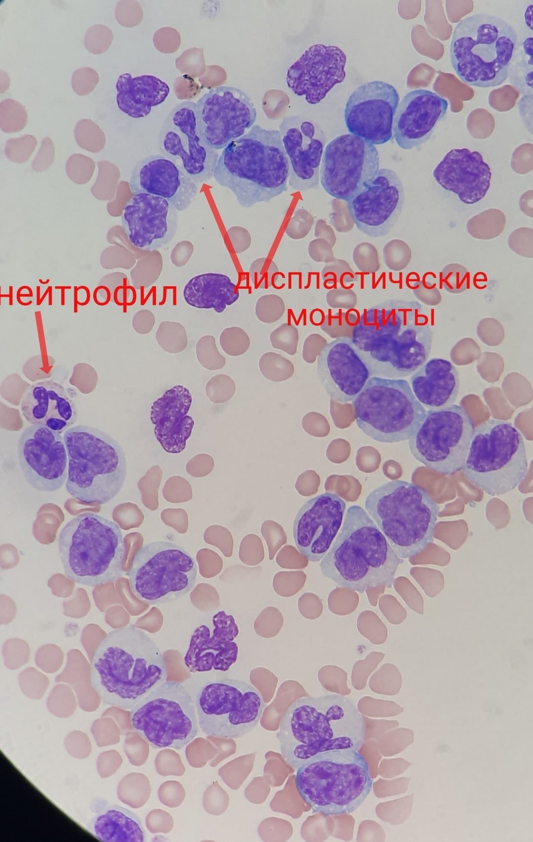
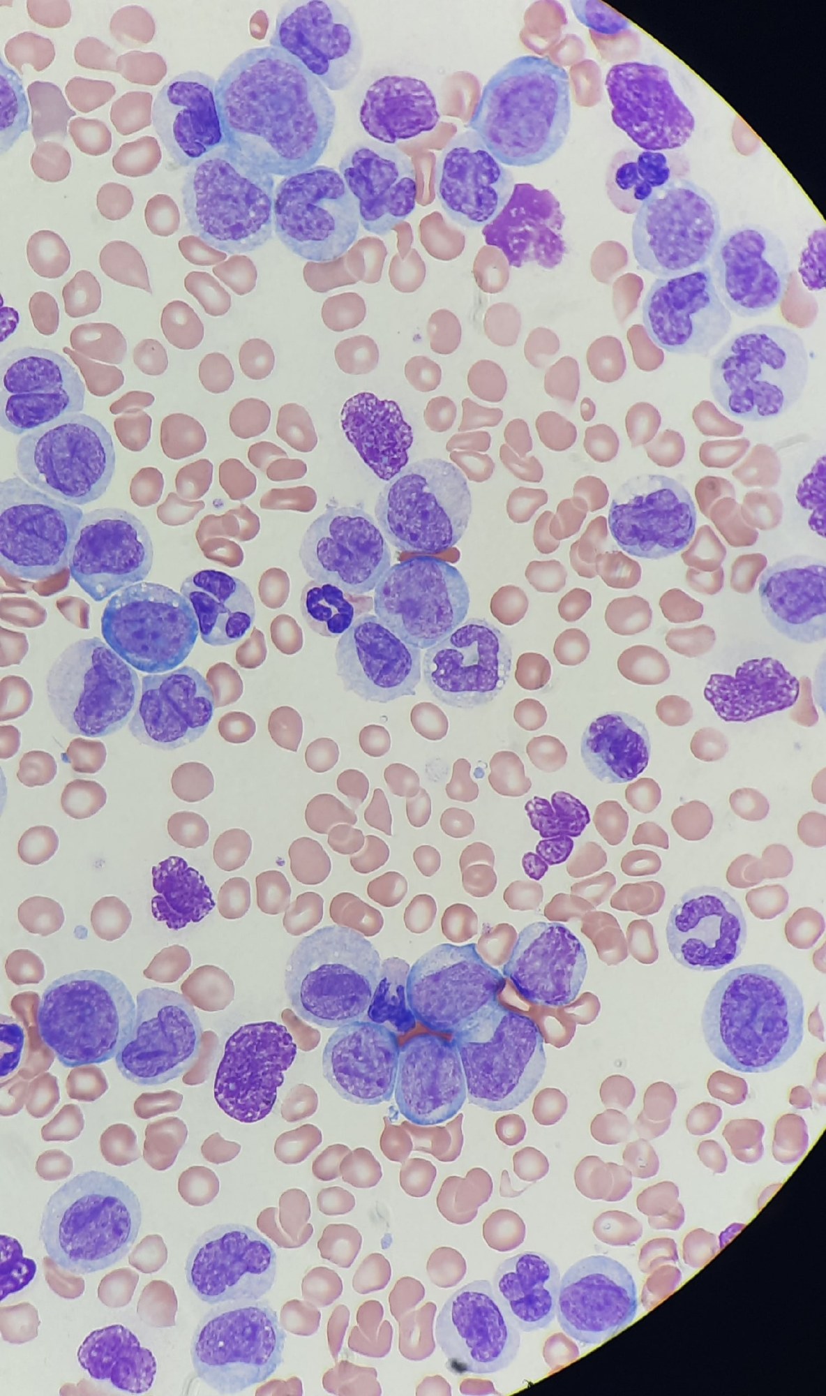
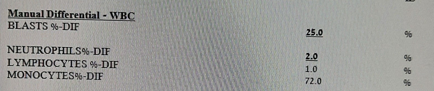
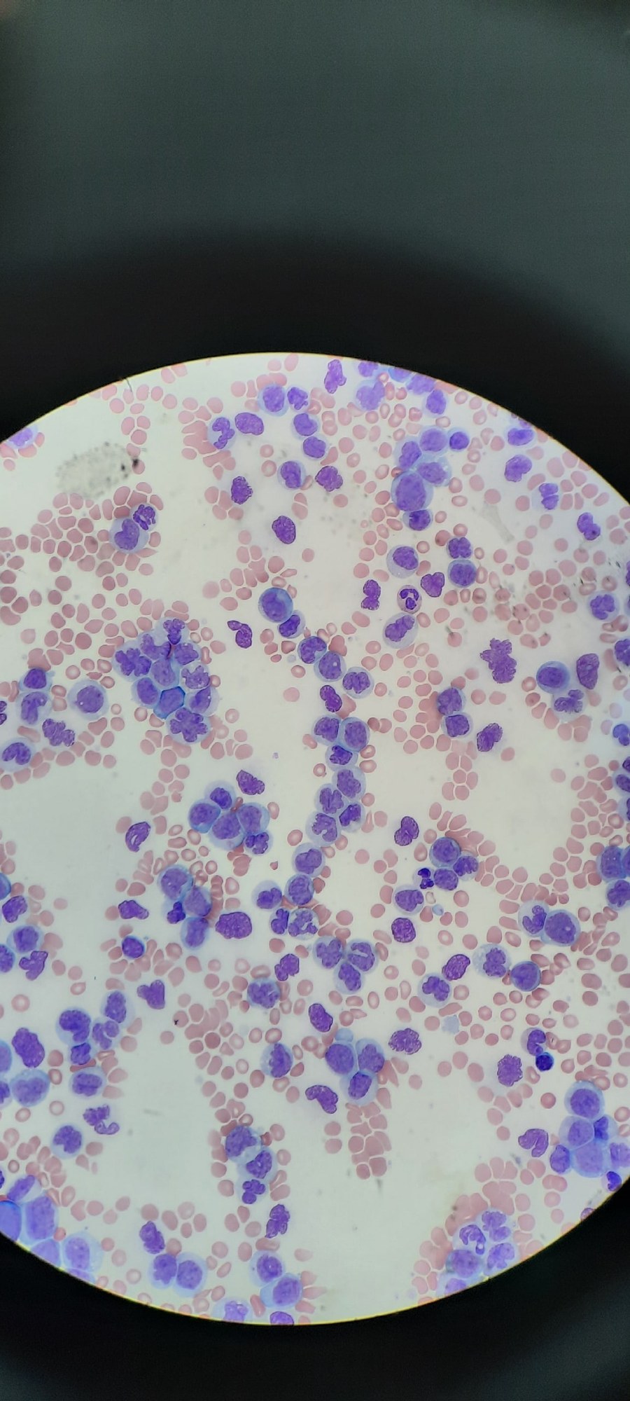
The smear shows hypercellularity, large cells with monocytic differentiation. Almost complete absence of granulocytes and lymphocytes. When counting under the microscope, I tried to identify monoblasts, and the remaining more mature cells were classified as monocytes with a description of immaturity and dysplasia (I didn’t bother with promonocytes). (See photo.)
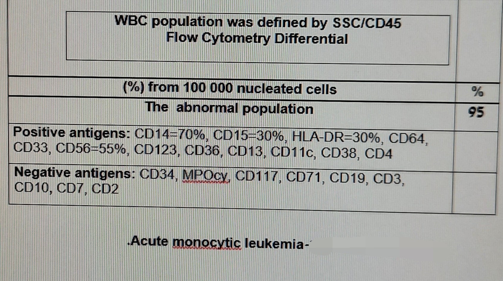
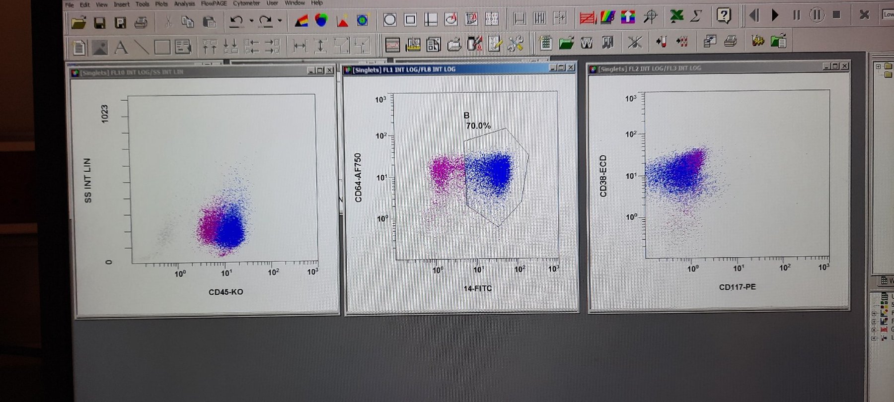
Immunophenotyping confirms monocytic origin, details of the phenotype (see photo) and the morphological picture lead to the conclusion: AML M5b (secondary, against the background of chemotherapy).
The patient received cytostatics; 4 days later, leukocytes dropped to 0.46×10⁹/L! Imagine such a sharp cell breakdown! LDH 931 (125-220), Creatine Kinase 2653 (30-200)!
12 days later, the patient died in the intensive care unit.


