

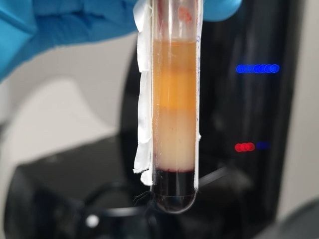
Did you know the difference between monocytic and monoblastic leukemia?
ACUTE MONOCYTIC LEUKEMIA
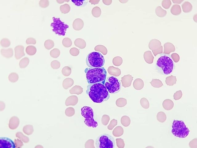
A 43-year-old female patient with complaints of general malaise, weight loss in recent weeks, and gum infiltration.
Complete blood count:
- Leukocytes: 340,000/µL
- Platelets: 17,000/µL
- Hemoglobin: 56 g/L
The result of the hematological blood test shows significant leukocytosis with anemia and thrombocytopenia, for which a peripheral blood smear is performed.
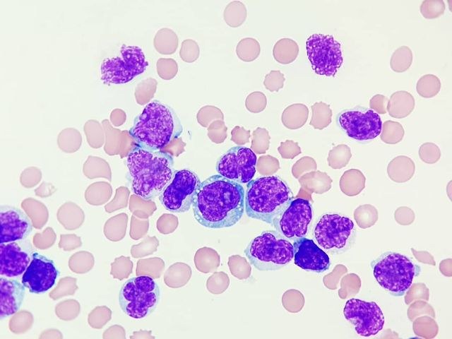
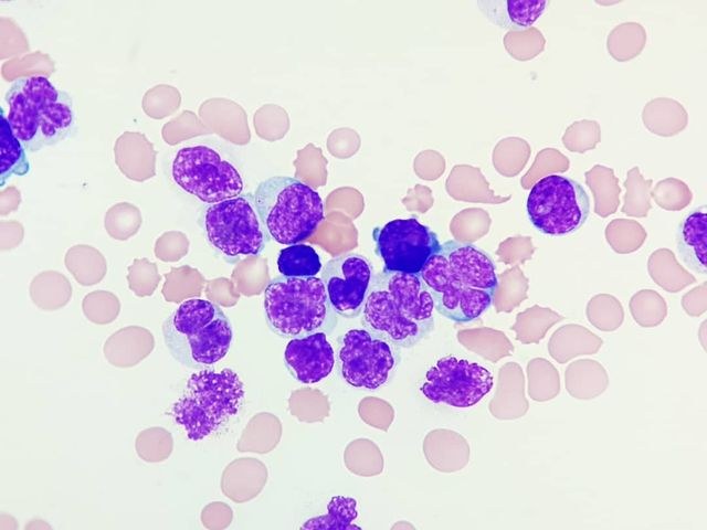
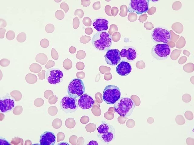
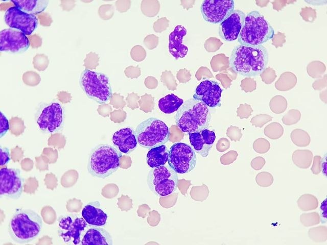
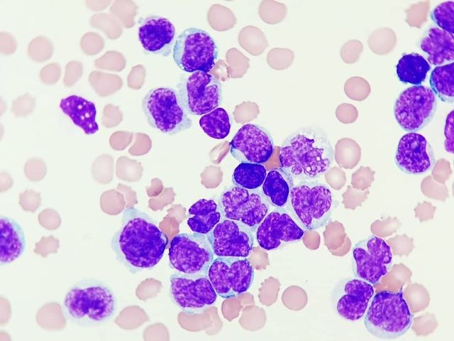
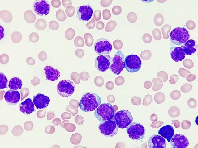
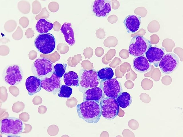
In the peripheral blood smear, cells are observed, some with fine chromatin and nucleoli (blasts), which constitute approximately 7% of the total cell count, while other cells have fine chromatin and convoluted nuclei, which together with monocytes constitute approximately 90% of the total cell count.
Based on the data obtained, flow cytometry is proposed.
Flow cytometry revealed a blast population, which constitutes approximately 5% of the total cell count, positive for CD34, CD117, HLA-DR, and CD64 antigens, and with weak expression of myeloperoxidase enzyme. In addition, a population of promonocytes and monocytes was found, which constitute approximately 86% of the total cell count.
Acute monoblastic leukemia and acute monocytic leukemia are myeloid leukemias in which blasts (including promonocytes) in peripheral blood or bone marrow constitute more than 20% and where more than 80% of tumor cells are of monocytic lineage, including monoblasts, promonocytes, and monocytes. The difference between them is based on the proportion of monoblasts and promonocytes in each; in acute monoblastic leukemia, more than 80% of monocytic cells are monoblasts, while in acute monocytic leukemia, most cells correspond to promonocytes or monocytes.
Source: inst:notashematologicas


