

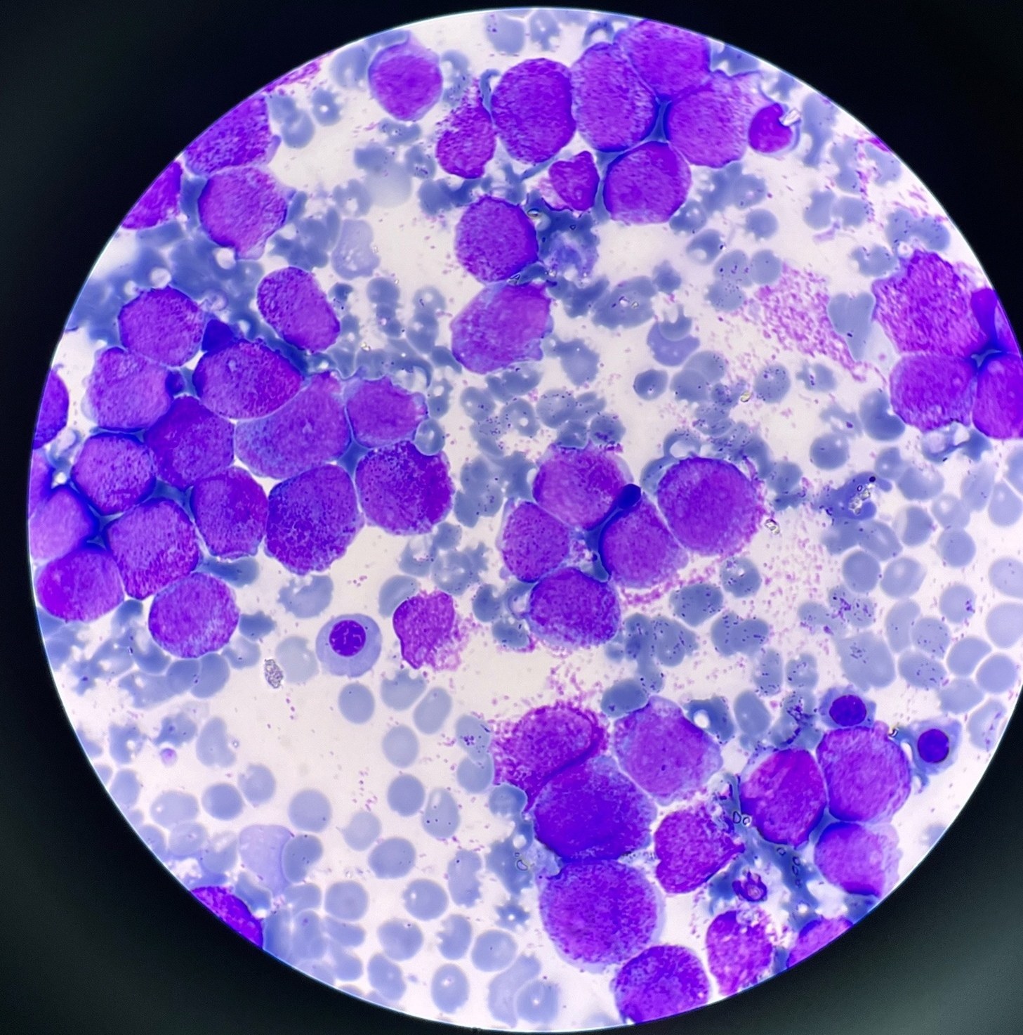
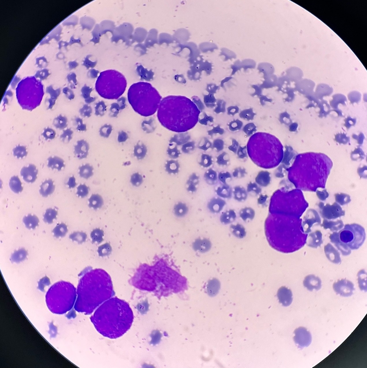
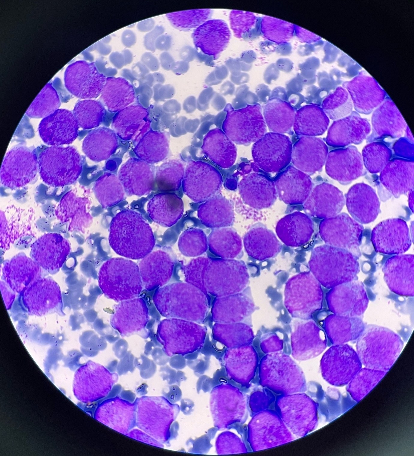
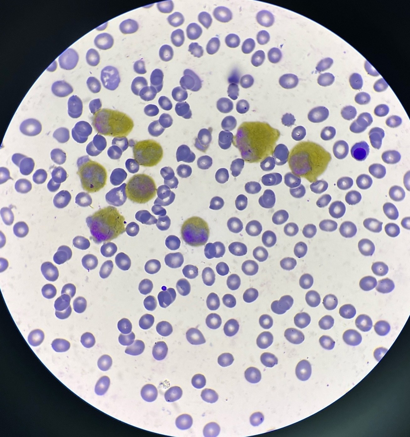
❗A task from the closed club of laboratory hematologists. ✅ A 56-year-old female patient, with pronounced hemorrhagic syndrome and weakness. She sought a hematologist herself. 🩸 Complete blood count: Leukocytes, WBC - 0.9 x 10³/µL ↓ Hemoglobin, HGB - 73 g/L ↓ Platelets, PLT - 8 x 10³/µL ↓ 🔬Microscopy of blood revealed 45% blasts. 🦴 Bone marrow picture in the images below (1-3 Romanowsky-Giemsa stain, 4 cytochemical stain for MPO). 💡Bone marrow immunophenotyping revealed a clone with the immunophenotype: CD45low, CD34-, HLA-DR-, CD123+low, CD117+, MPO+, CD33+, CD64+low, CD13+, CD15-, CD11b+low, CD61-, CD235a-, cytCD3-, CD7-, CD3-, CD8-, CD4+low, cytCD79a-, CD19-, CD20+low, CD10-. ❓1. What diagnosis can we assume? 2. Which chromosomes should be investigated in cytogenetics? 3. Can we prescribe treatment now? ⭐ The first 7 people who correctly answer these 3 questions in the comments will receive an invitation to the closed club next week.
👉 The task was kindly prepared by Adelina Valeryevna Polyakova, a clinical laboratory diagnostics physician, Barnaul, Regional Clinical Hospital.


