

Clinical case: Acute B-lymphoblastic leukemia with MEF2D::BCL9 rearrangement mimicking Burkitt lymphoma.
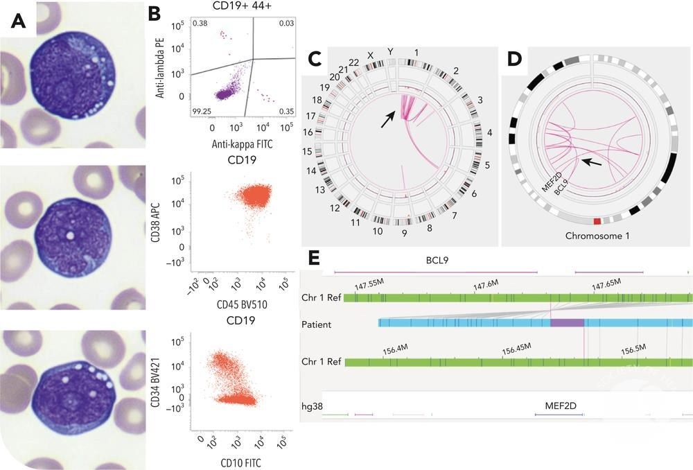
A 39-year-old female patient presented with severe lymphadenopathy, leukocytosis (25.1 × 10⁹/L), anemia (hemoglobin 96 g/L), thrombocytopenia (23 × 10⁹/L), and significantly elevated lactate dehydrogenase activity (8594 U/L). Peripheral blood smear revealed 46% atypical cells with finely clumped chromatin, medium to large nucleoli, basophilic cytoplasm, and prominent cytoplasmic vacuoles, morphologically resembling Burkitt lymphoma cells (panel A: May-Grünwald-Giemsa stain, ×1000 magnification).
Massive bone marrow infiltration was confirmed morphologically. Immunophenotyping revealed a population of immature B-cells, positive for CD19, cCD79a, CD22, CD10, CD38, terminal deoxynucleotidyl transferase, and CD34 (on a subset of cells), but negative for CD20, as well as surface and cytoplasmic immunoglobulins (panel B).
Fluorescence in situ hybridization did not detect MYC and BCR::ABL1 rearrangements. Cytogenetic analysis showed a complex karyotype: 46,XX,der(1)(2pter→2p11::1p31→1q21::?::2q13→2qter),-2,-6,+2mar[20]. Optical genome mapping revealed a pattern of chromothripsis on chromosome 1 (Circos plots in panels C and D [indicated by arrow]), including a MEF2D::BCL9 rearrangement (panels D and E). The rearrangement was confirmed by targeted RNA sequencing.
❗Key points: Morphology: Leukemia with MEF2D::BCL9 rearrangement morphologically mimics Burkitt lymphoma cells, which can lead to diagnostic difficulties.
Cytogenetics and molecular diagnostics: This case highlights the importance of using cytogenetic and molecular methods, such as optical genome mapping and targeted RNA sequencing, for accurate diagnosis.
Clinical significance: Acute lymphoblastic leukemia with MEF2D rearrangement is classified as a distinct subcategory in the International Classification of Tumors and the 5th edition of the WHO classification. These forms of leukemia are associated with high risk, making their identification critically important for selecting the optimal therapeutic approach.
Conclusion: The presented case demonstrates the necessity of a comprehensive approach to the diagnosis of acute lymphoblastic leukemias. Morphological features of the cells combined with molecular-cytogenetic methods allow for the identification of rare and highly aggressive subtypes, which plays a crucial role in patient prognosis and treatment.
👉 Source: imagebank hematology. Published Date: 11/13/2024 Author: Simon Chevalier; Clémentine Legrand


