

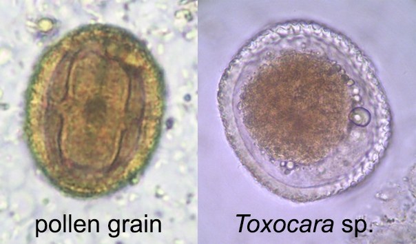
✅ANSWER to the parasitology quiz: Pollen grains.
🎊Congratulations to everyone who provided the correct answer.
✅These structures externally resemble ascarid eggs. Size and surface texture will help differentiate them. Toxocara canis measures 80-85 micrometers in its greatest dimension, and its surface texture resembles a golf ball. T. cati also measures 65-70 micrometers, has a golf ball-like pitting, but is smaller and less distinct than T. canis. Baylisascaris procyonis measures 63-88 micrometers and has a granular surface texture. Always check the surface texture by focusing up and down on the focal plane of the egg’s surface. It’s amazing what you will see. This is how a mixed infection of T. canis and B. procyonis was once diagnosed in a dog. In the photo below: a parallel comparison of pollen in our case and a Toxocara sp. egg. Also note the pores, which help distinguish a pollen grain from a true helminth egg.
✅It might be cornflower (Psephellus) pollen. We don’t know much about pollen, but a quick Google search revealed some good photos of several pollen genera that, in this case, bear a resemblance to cornflower pollen.
📢Want another quiz?


