

Image of a blood smear from a patient with cryoglobulinemia, showing a large extracellular cryoglobulin deposit.
Source: Bain Barbara J. - Hematology - 101 Morphology Updates, 2023
Morphological features of a peripheral blood smear in cryoglobulinemia: large extracellular amorphous protein deposit among red blood cells.
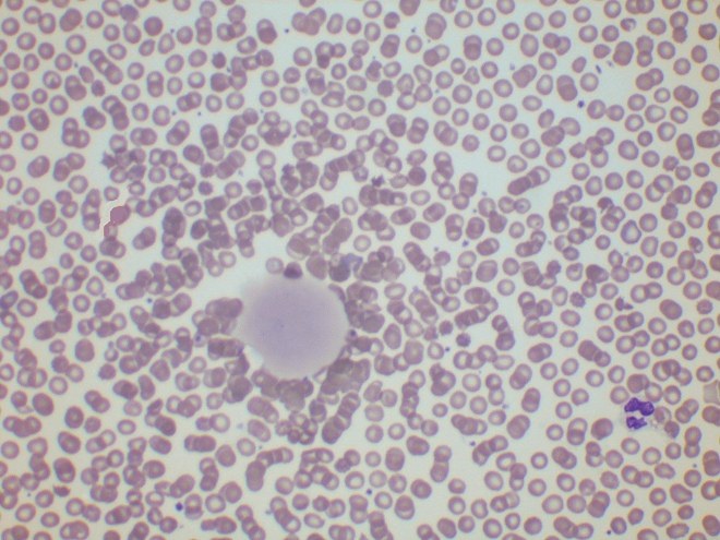
Image of a blood smear from a patient with cryoglobulinemia, showing a large extracellular cryoglobulin deposit.
Source: Bain Barbara J. - Hematology - 101 Morphology Updates, 2023

Laboratory hematology test: identify the type of granulocytes with dermatan sulfate inclusions in a peripheral blood smear using a photomicrograph.

Analysis of rare diagnostic findings in blood and bone marrow smears: plasmacytes, schistocytes, erythrophagocytosis, blasts, and malaria trophozoites.

A diagnostic exercise in blood smear microscopy: identify the leukocytes presented in a routine blood test from a patient without specific complaints.