

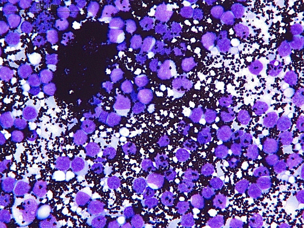
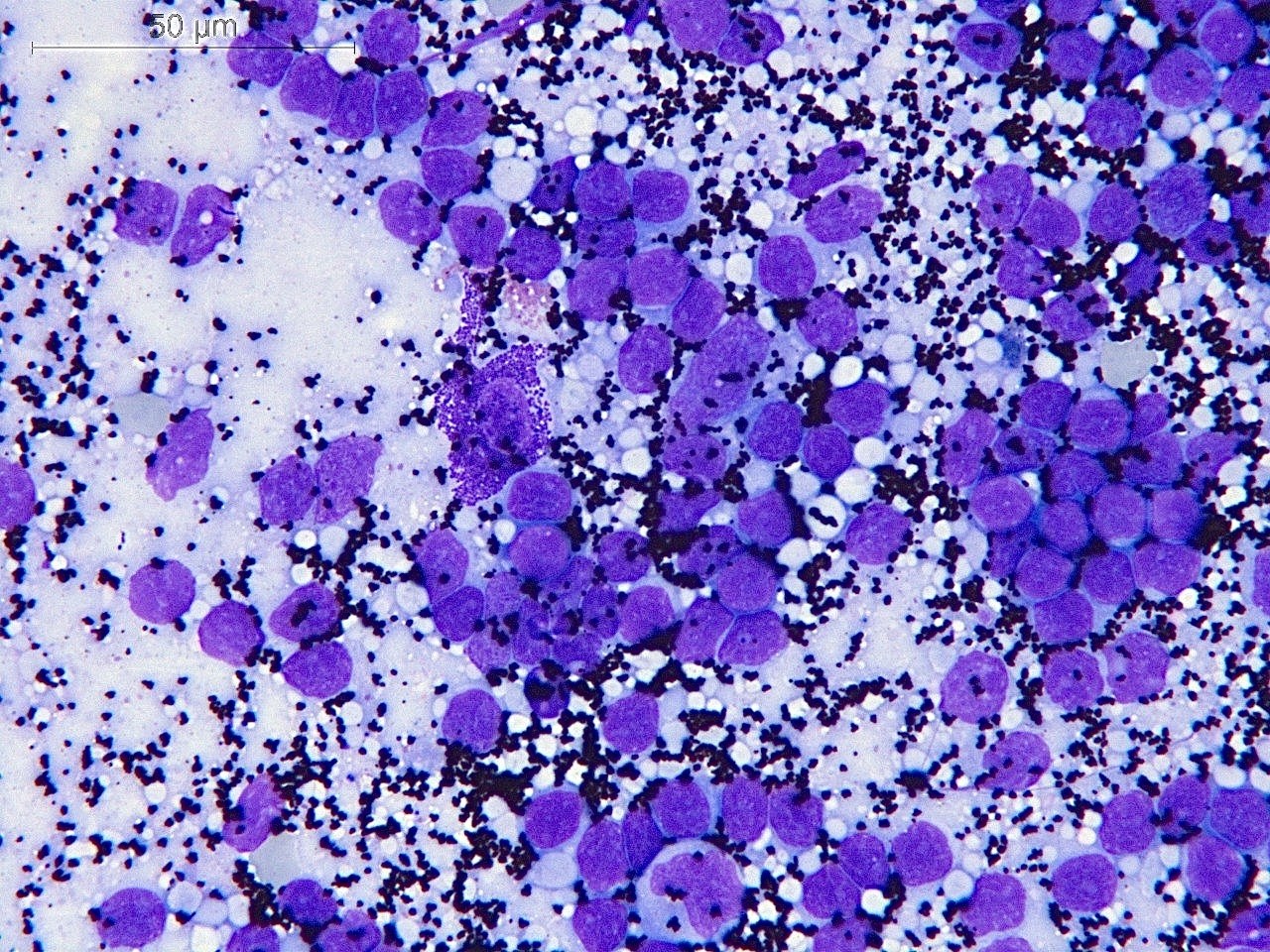
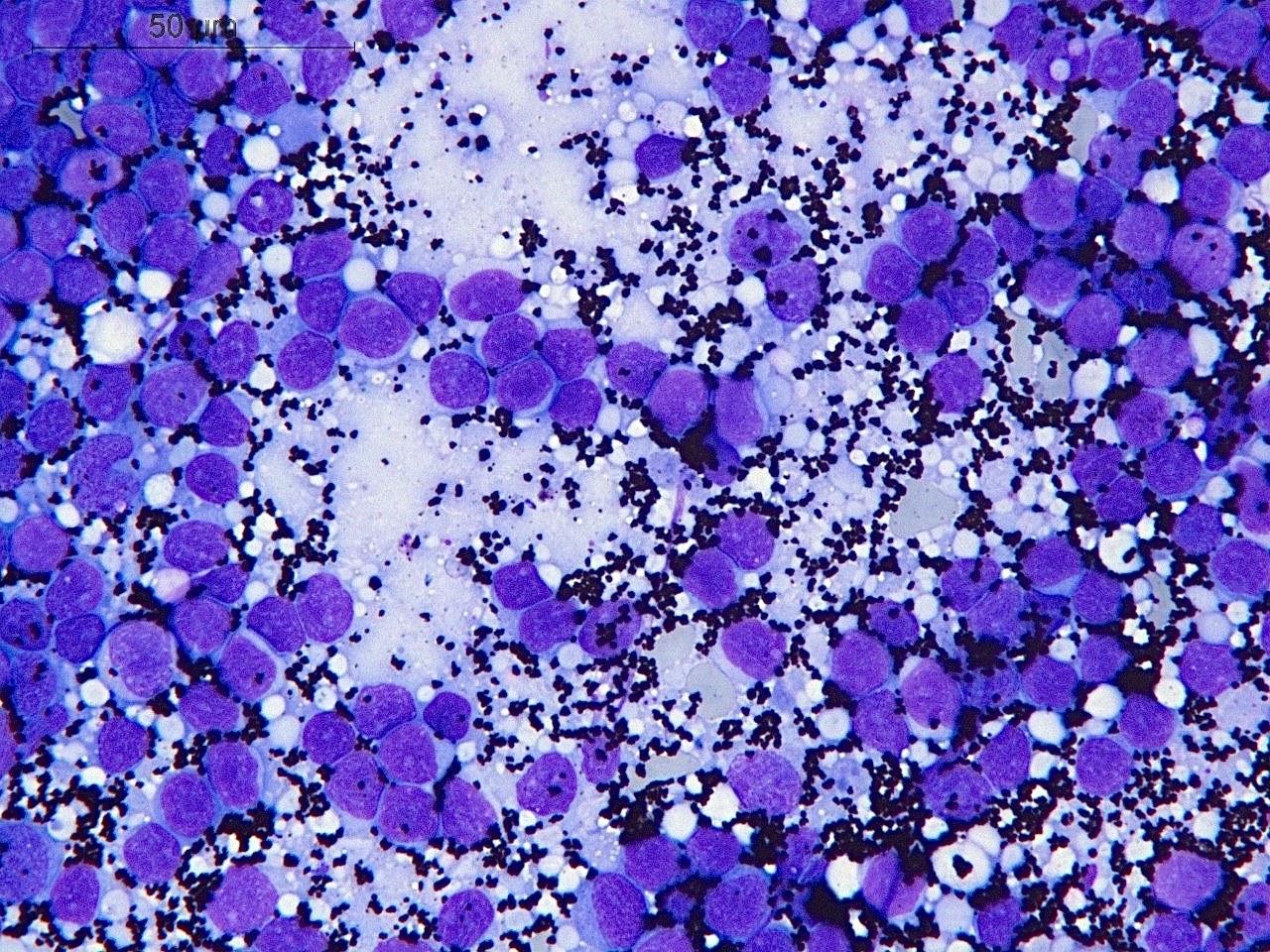
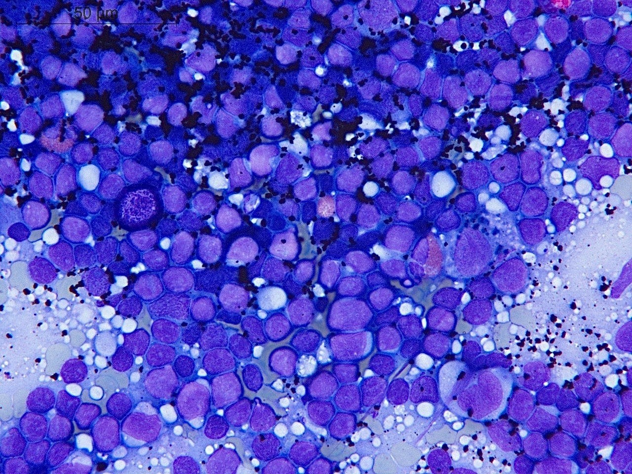
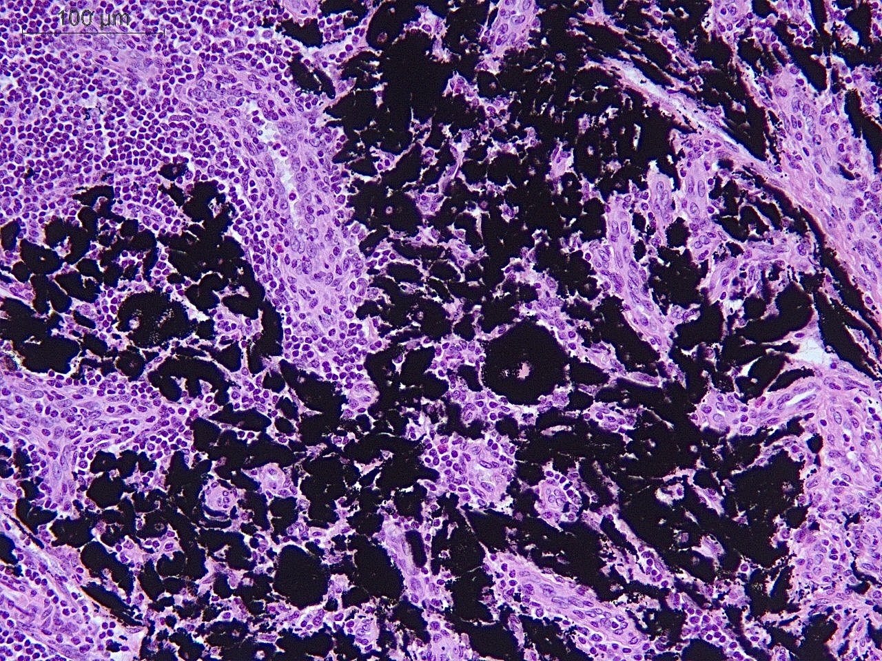
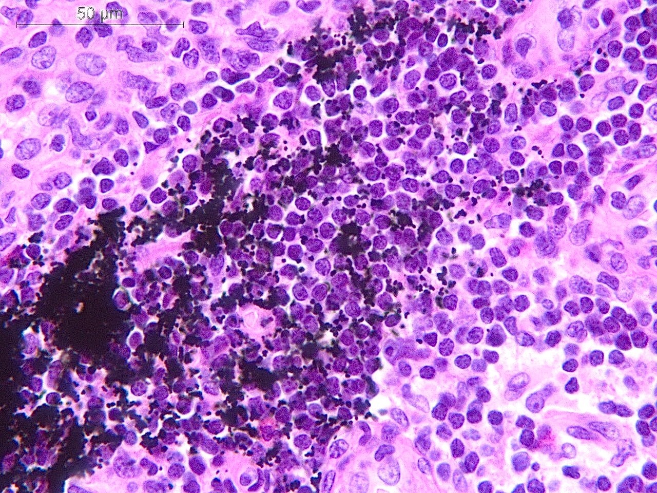
🔬 Inguinal lymph node of a 22-year-old woman. Photos of cytological and histological preparations show massive deposits of black pigment. The pigment is located both interstitially and in the cytoplasm of histiocytes. No tumor growth was detected. The observed changes correspond to exogenous pigmentation (tattoo pigment). Be careful with tattoos, especially colored ones!
🙏 The case was kindly provided by Clinical Laboratory Diagnostics Doctor, Pathologist, member of the FLM Hematology Committee Elena Belyakova, Department of Molecular Morphology, E.E. Eichwald Clinic, I.I. Mechnikov Northwestern State Medical University, St. Petersburg.


