

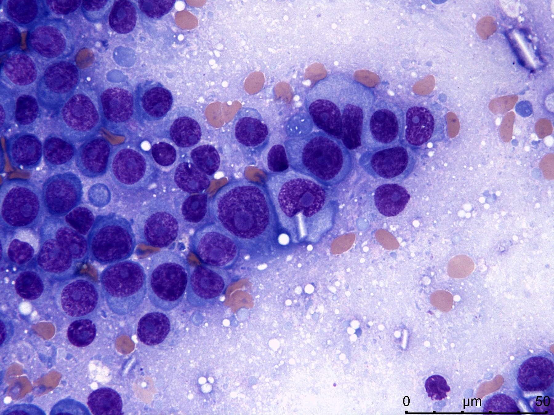
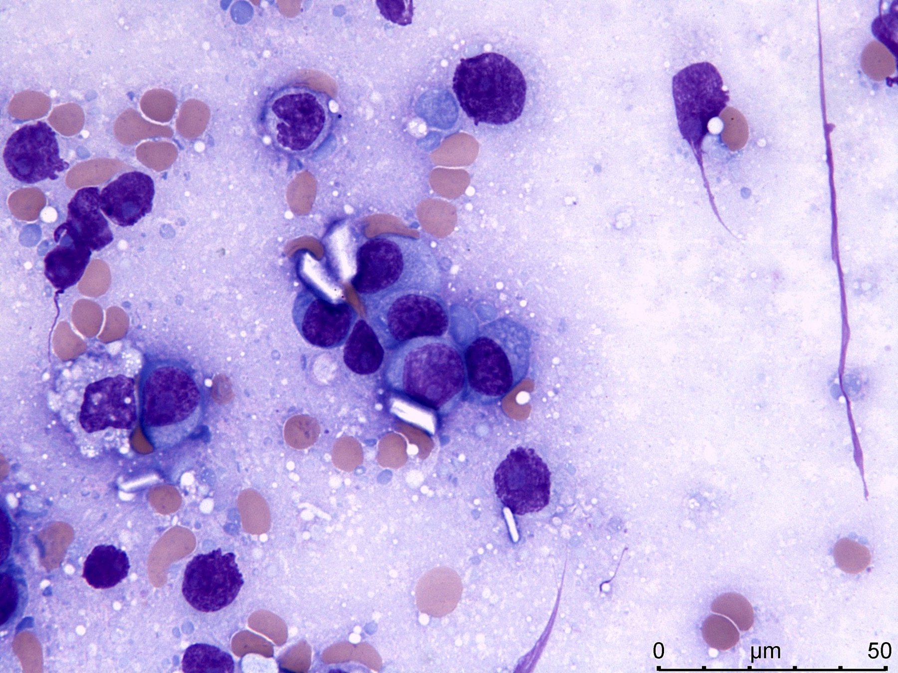
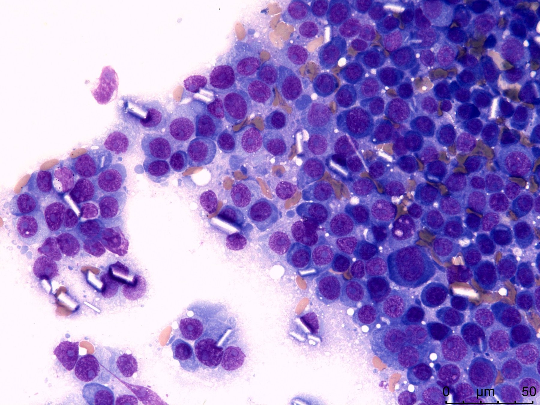
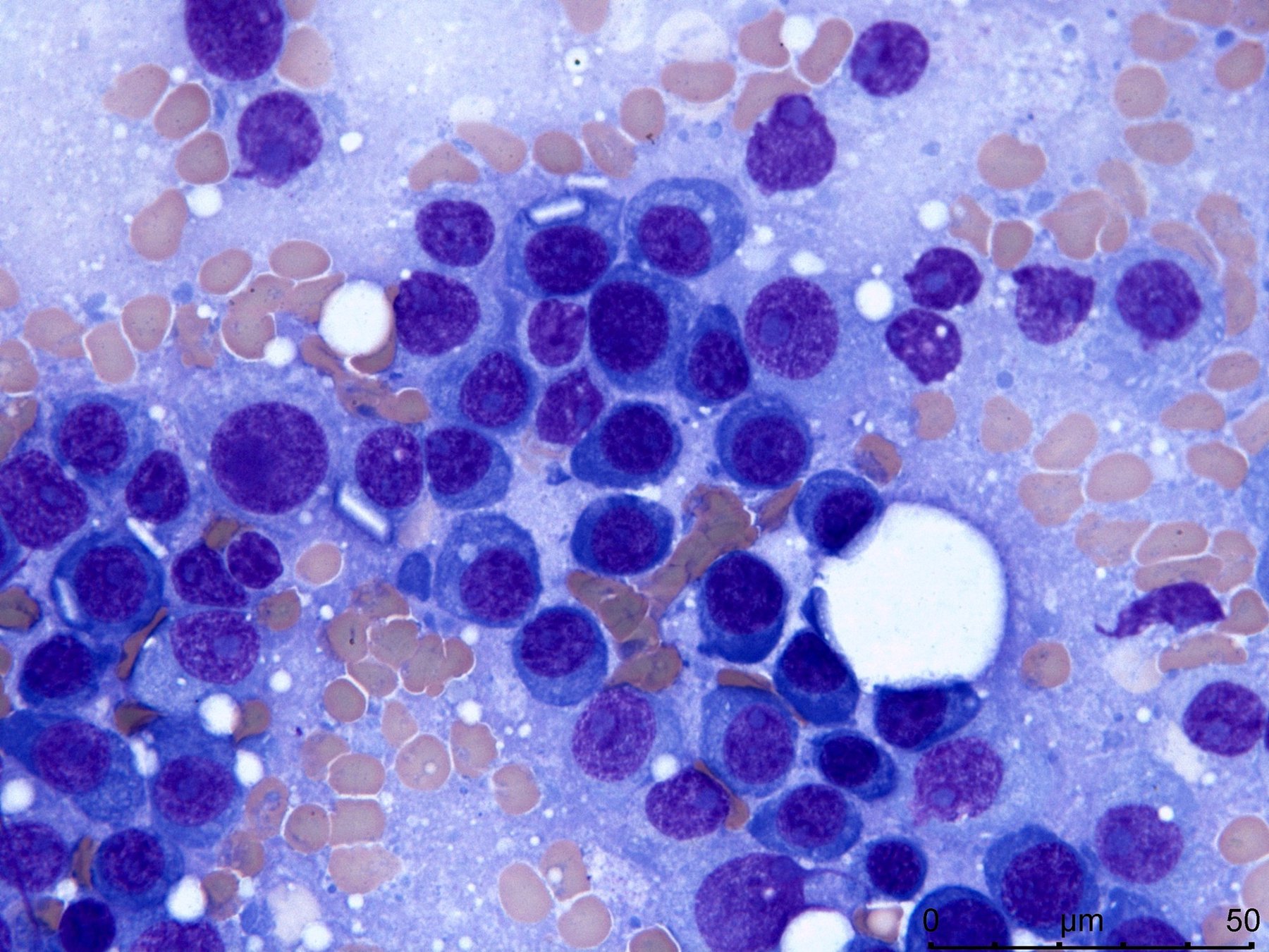
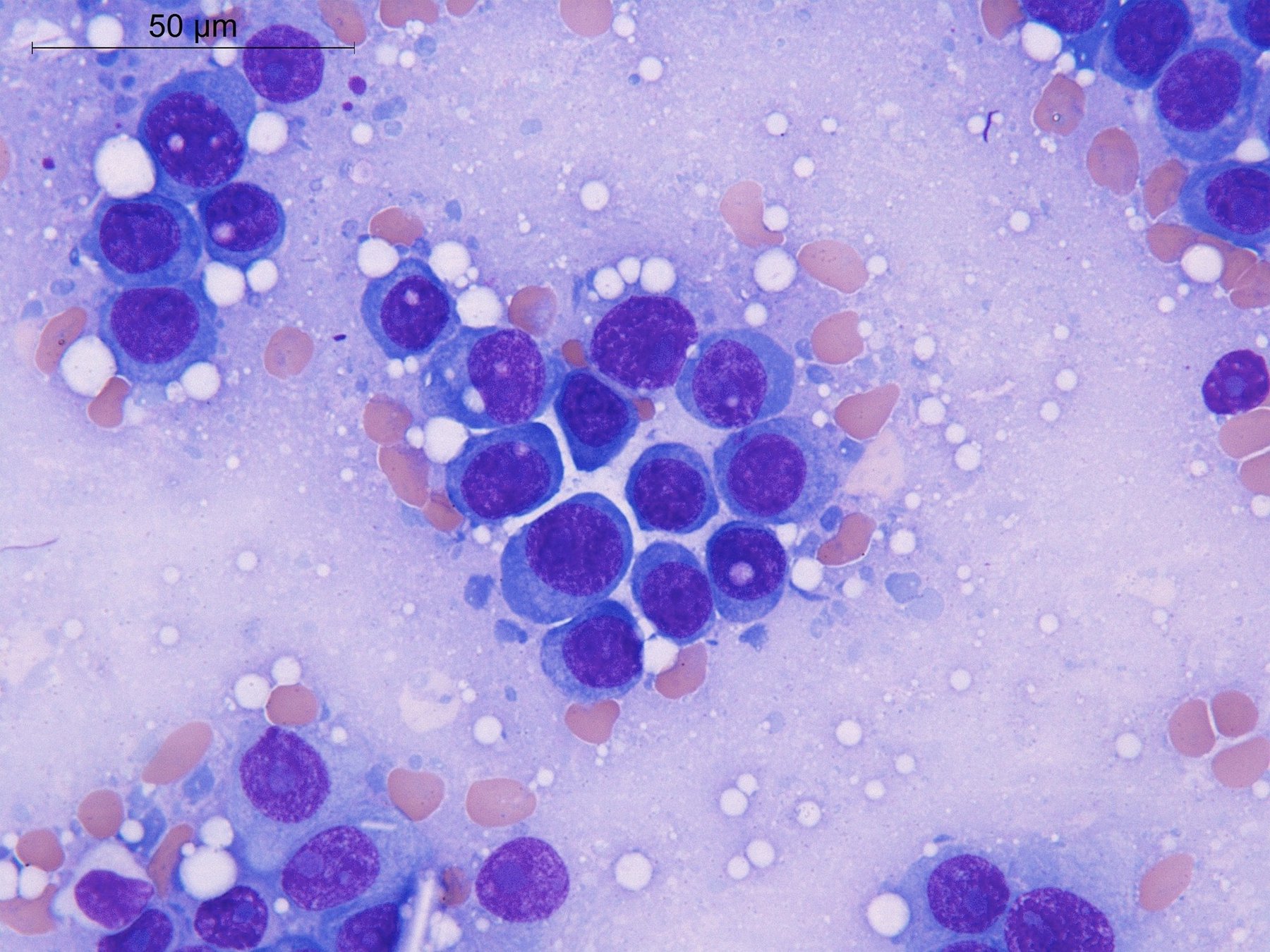
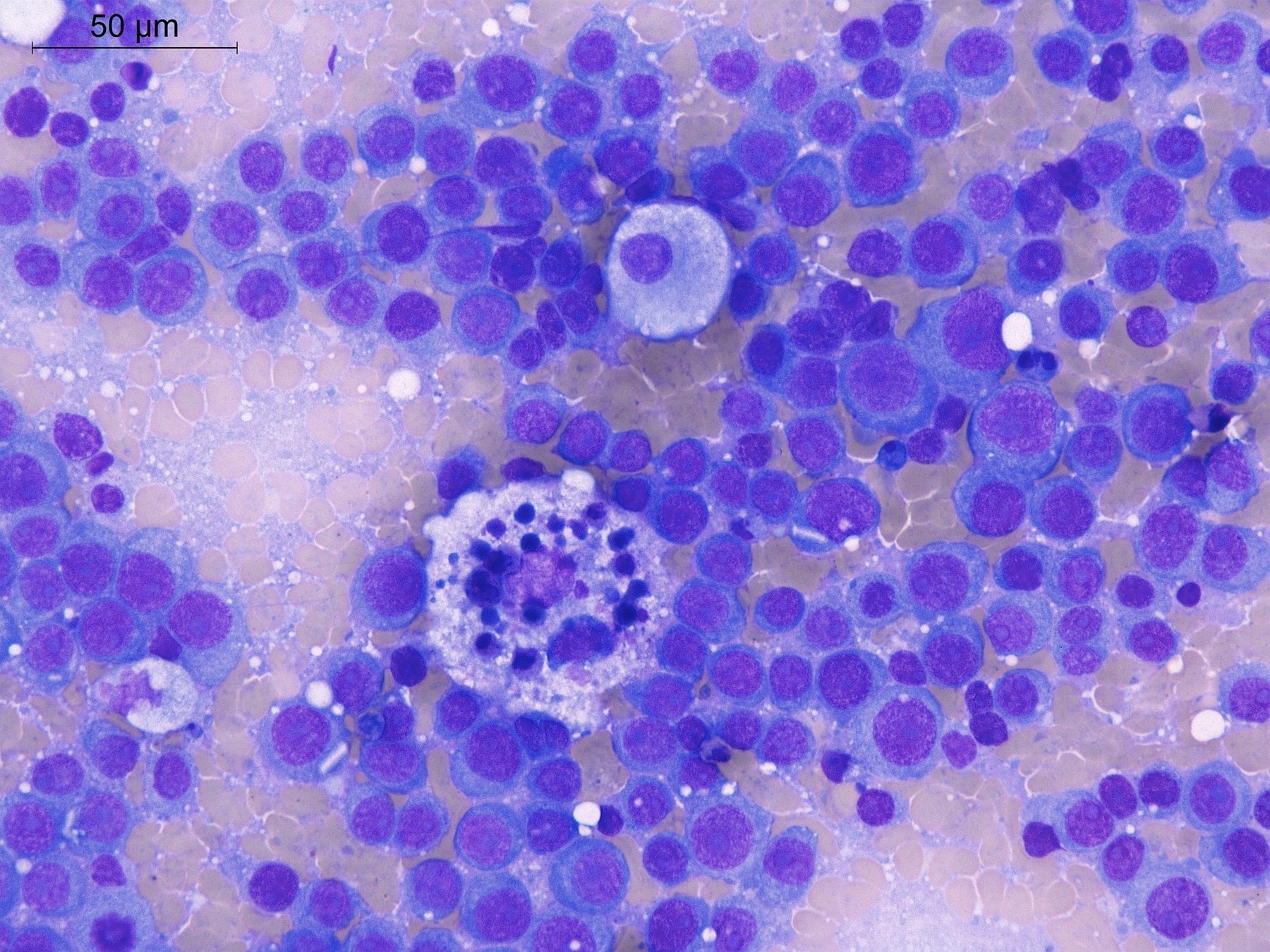
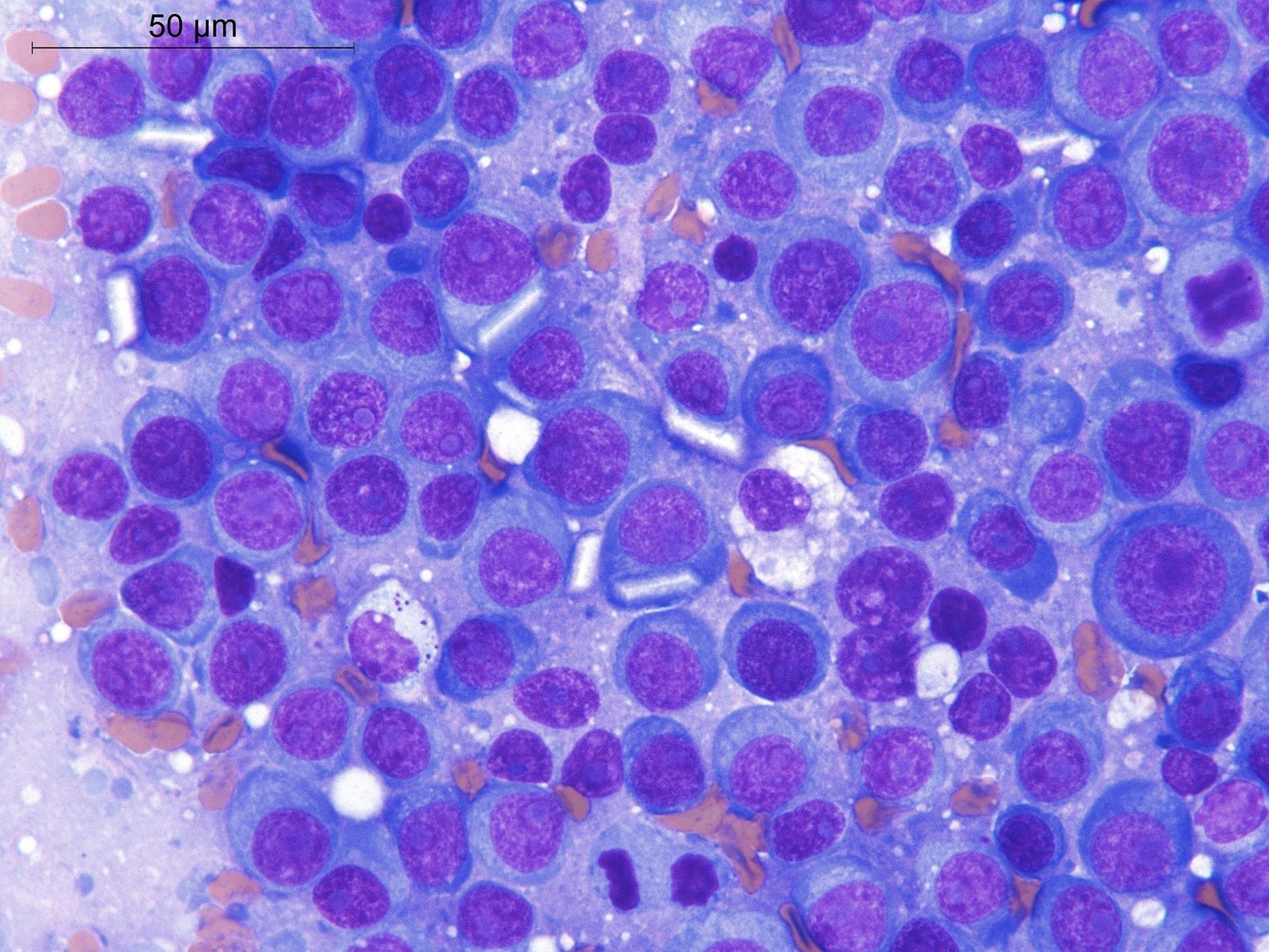
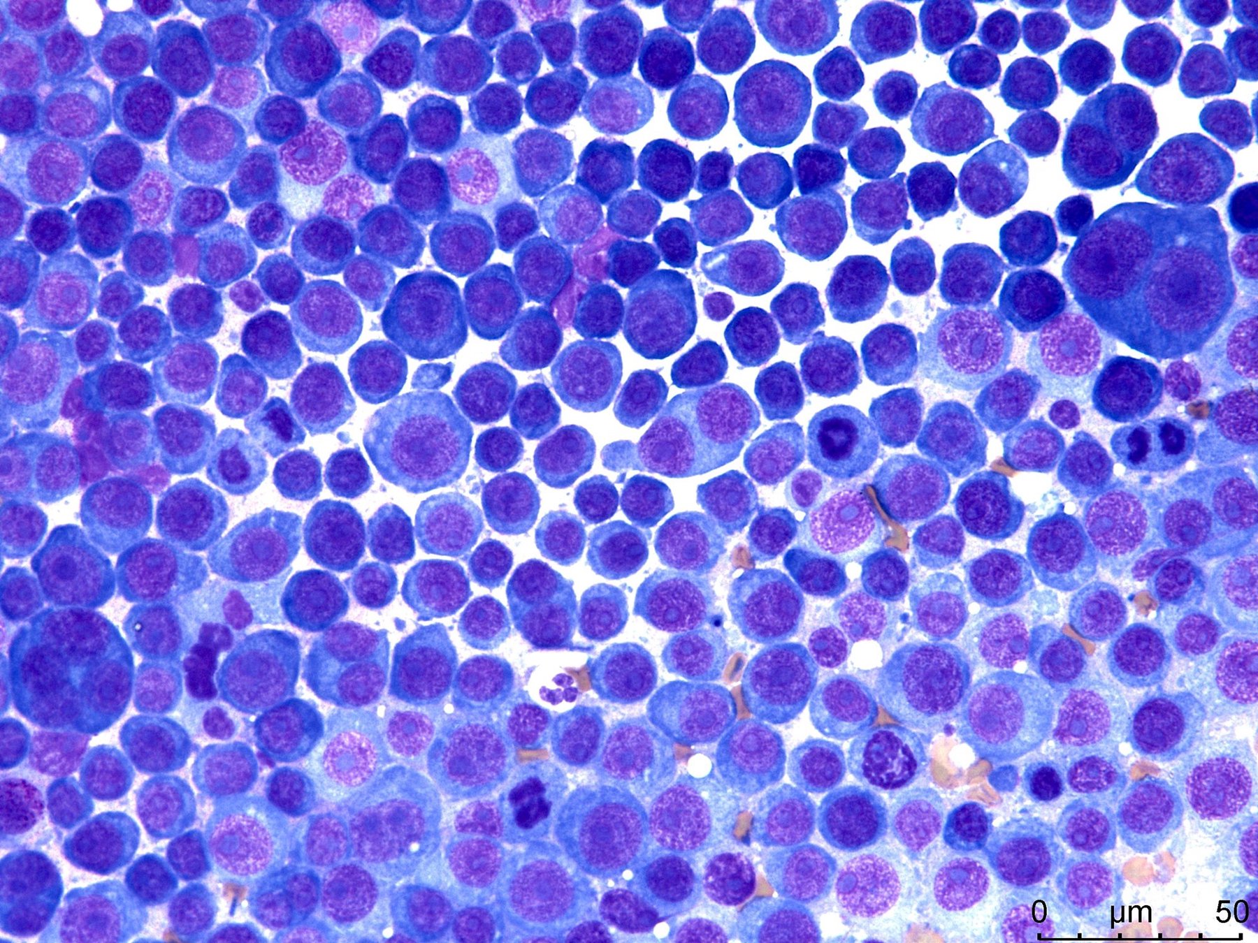
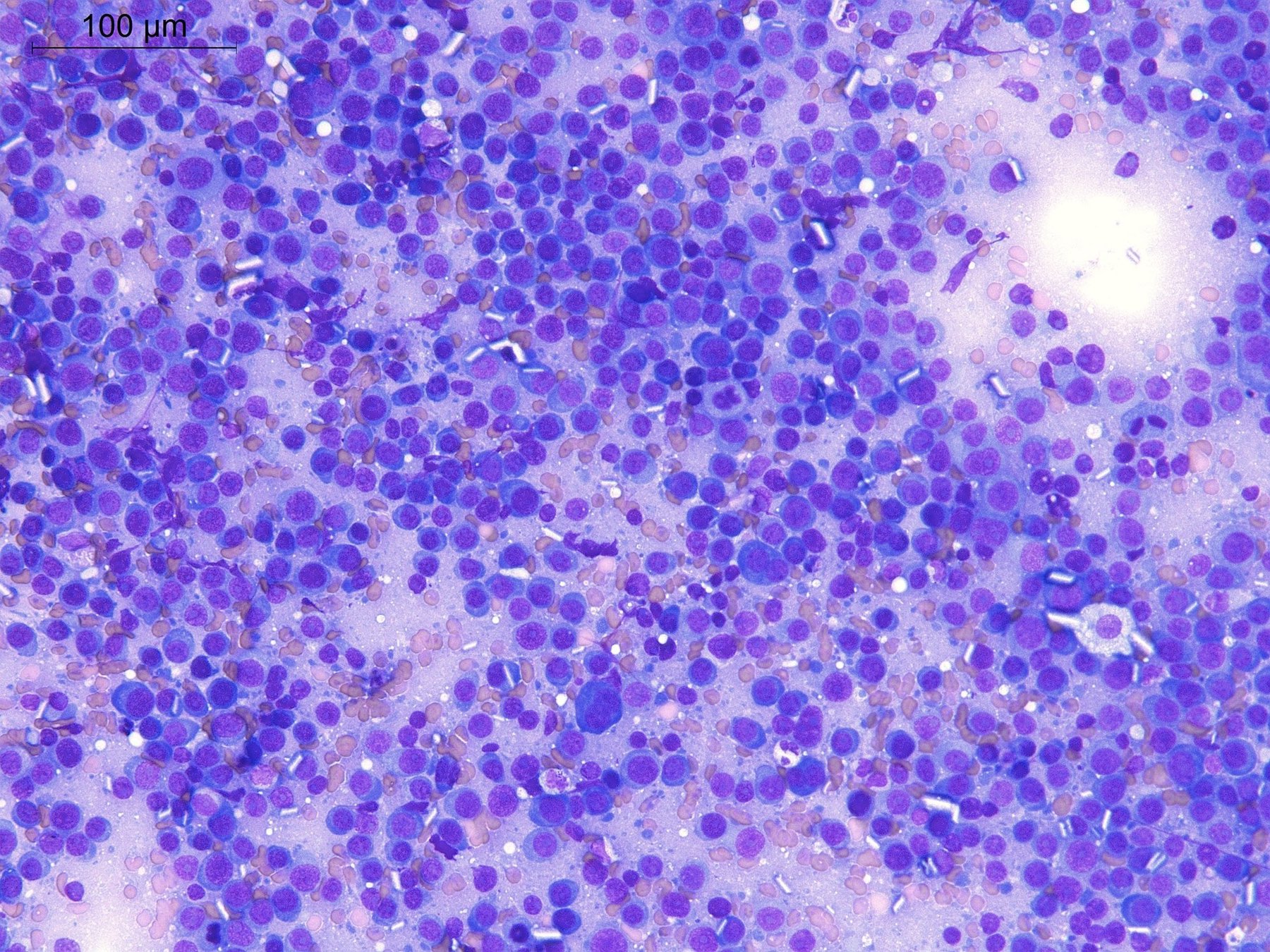
🔬 Lymph node imprint from a patient with a previously diagnosed multiple myeloma. Progression occurred during chemotherapy. The photographs show fields of plasmacytes and plasmablasts, with crystalline immunoglobulin inclusions in the cell cytoplasm, as well as free-lying crystals. The proliferative activity index Ki-67 of these tumor cells is close to 100% according to IHC data. This is how plasmacytoma (plasmablastic transformation of multiple myeloma) appears.
🙏 The images and description were kindly provided by clinical laboratory diagnostics physician, pathologist, member of the FLM Hematology Committee Elena Belyakova, Department of Molecular Morphology, E.E. Eichwald Clinic, I.I. Mechnikov Northwestern State Medical University, St. Petersburg.


