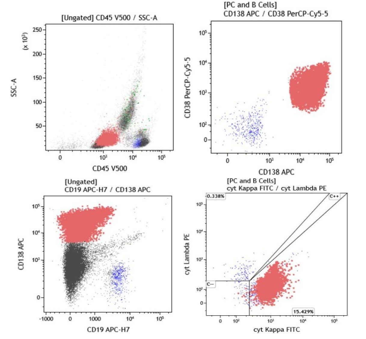
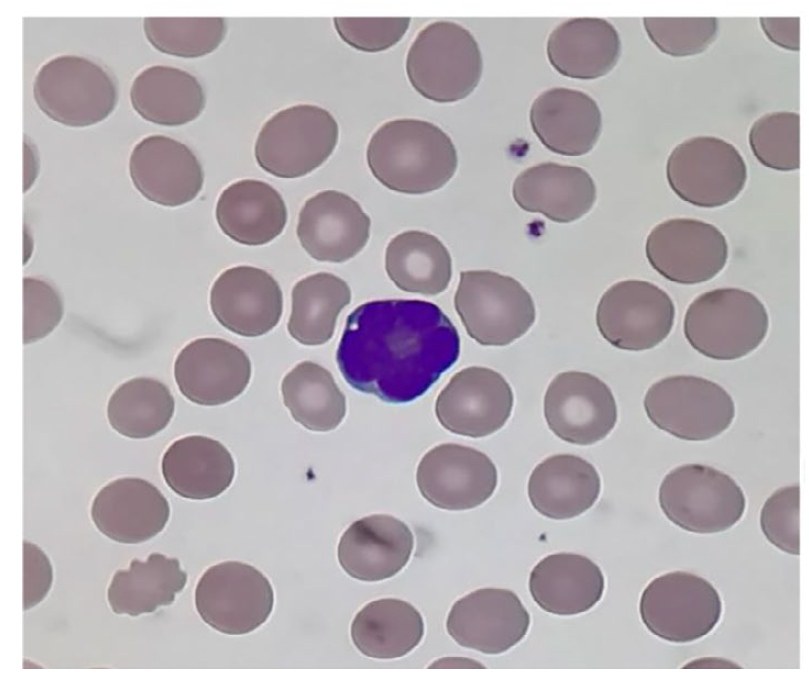
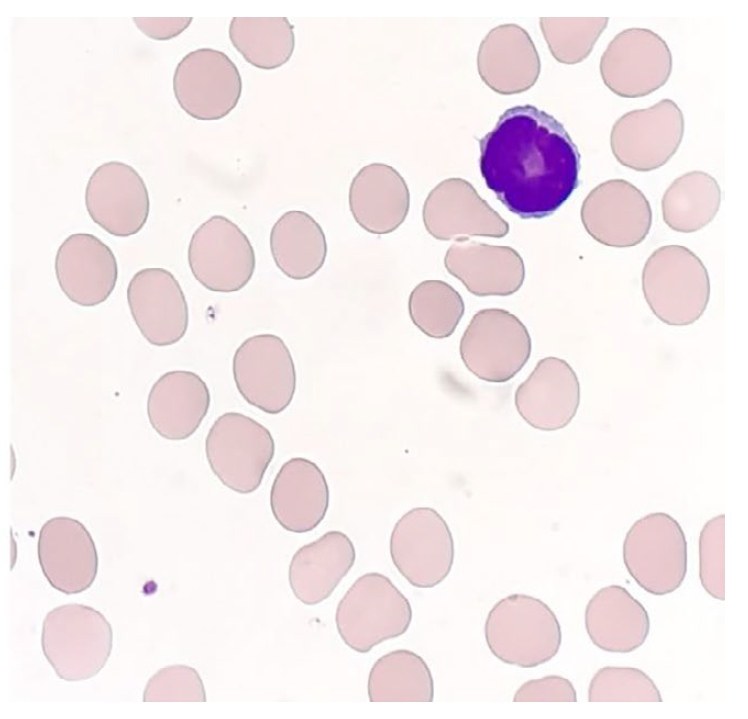
PLASMA CELL LEUKEMIA WITH “LYMPHOID-LIKE MORPHOLOGY” IN PERIPHERAL BLOOD. A 59-year-old man with a history of hypertension presented to his doctor with a 1-week history of lower urinary tract symptoms, for which he was prescribed a 7-day course of nitrofurantoin. Given the lack of improvement and the appearance of new non-specific symptoms of malaise, a series of investigations were required. ✅EXAMINATION Physical examination was unremarkable. ✅COMPLETE BLOOD COUNT: High white blood cell count (WBC) 43.3 × 10⁹/L, Hemoglobin 104 g/L, Platelet count 160 × 109 / L. The automated cell counter (Sysmex XE 2100®, Sysmex Corp) noted lymphocytosis and atypical lymphocytes in the sample. ✅BLOOD SMEAR MICROSCOPY Peripheral blood (x40, May-Grünwald-Giemsa stain). A monomorphic population of medium-sized mononuclear “lymphoid” cells with abnormal nuclear convolutions, prominent nucleoli, and scanty basophilic cytoplasm, comprising the majority of circulating leukocytes (photo below). ✅BIOCHEMISTRY In addition to the abnormalities in the complete blood count, the patient had acute kidney injury with creatinine 377 µmol/L (62-106) and urea 19.9 mmol/L (2.5-7.8), hypercalcemia with corrected calcium 3.99 mmol/L (2.2-2.6). Lactate dehydrogenase level was normal. ✅COMPUTED TOMOGRAPHY (CT) Computed tomography of the neck, chest, pelvis, and abdomen without contrast revealed no enlarged lymph nodes or hepatosplenomegaly; rather widespread osteolytic lesions were noted. ✅IMMUNOPHENOTYPING Peripheral blood immunophenotyping identified abnormal clonal plasma cells, comprising 66% of total nucleated cells (28.6 × 10^9/L). These cells were positive for CD 38, CD 138, and cytoplasmic kappa and were negative for CD 45, CD 56, and CD 19 (photo below). ✅PROTEIN ELECTROPHORESIS Accordingly, serum protein electrophoresis was requested, but no monoclonal band was detected, but hypogammaglobulinemia was present. Serum-free light chain analysis showed a kappa free light chain count of 3654.8 mg/L (3.3-19.4) with a kappa/lambda ratio of 589.48. Urine protein electrophoresis identified a free monoclonal kappa light chain band, comprising 76.4% of total urine protein and quantified at 0.44 g/L. ✅CYTOGENETICS Cytogenetic studies revealed a variant translocation t (11;14) (q13;q32). ✅FINAL DIAGNOSIS Primary plasma cell leukemia. ✅PROGNOSIS Primary plasma cell leukemia is a rare and aggressive malignancy. ✅SOURCE Nesr, Maniusyte, R., Dunlop, A., & Kagdi, H. (2021). Plasma cell leukaemia with “lymphoid‐like morphology” in peripheral blood. International Journal of Laboratory Hematology, 44(1), 10–12. Portico. ✅OUR CONCLUSIONS Immunophenotyping is the gold standard in diagnosis.
Primary Plasma Cell Leukemia with Lymphoid-Like Morphology
A case study of primary plasma cell leukemia with lymphoid-like cell morphology: blood smear, flow cytometry, and cytogenetics data.