

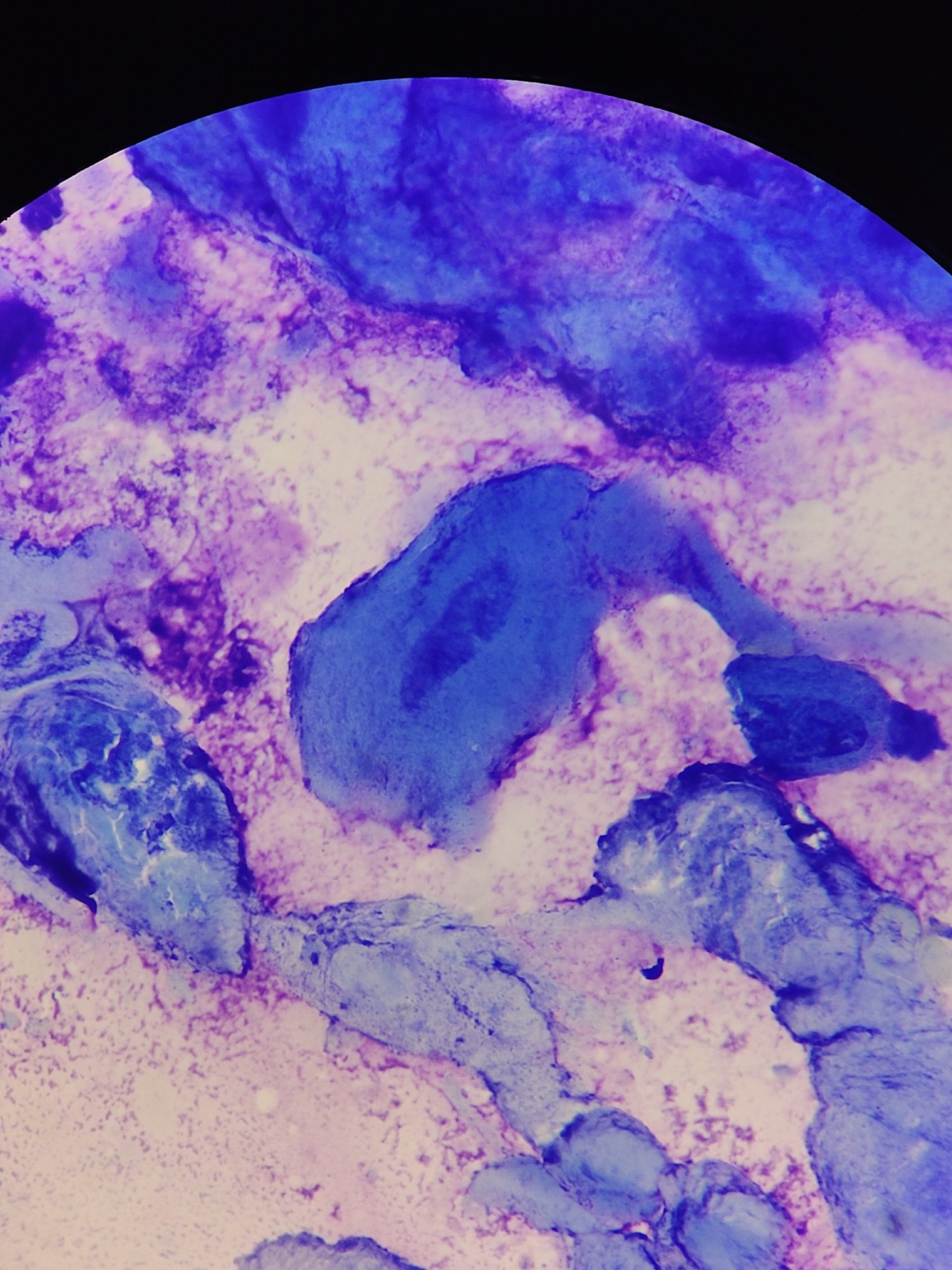
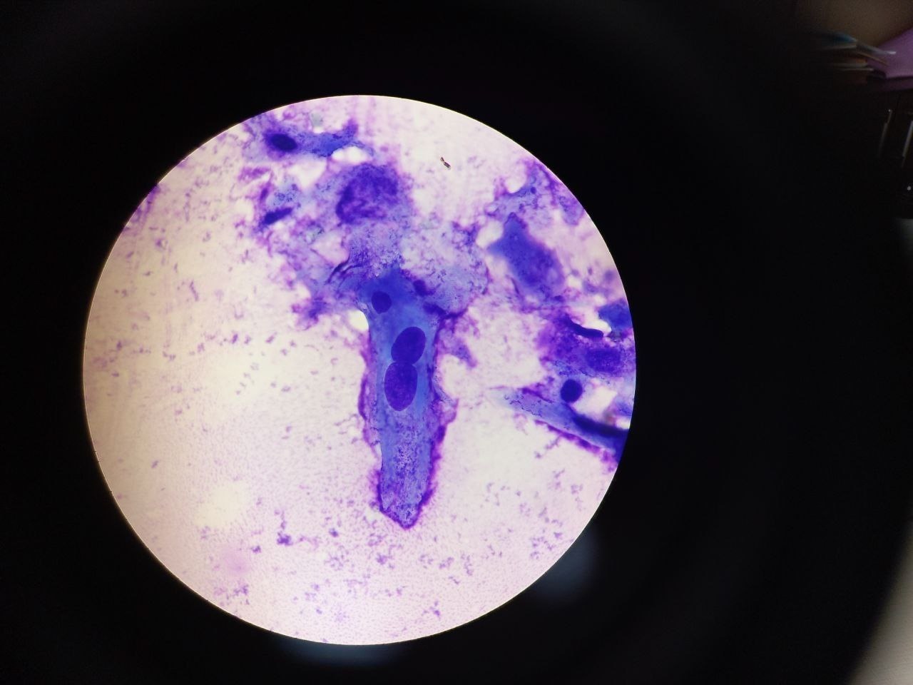
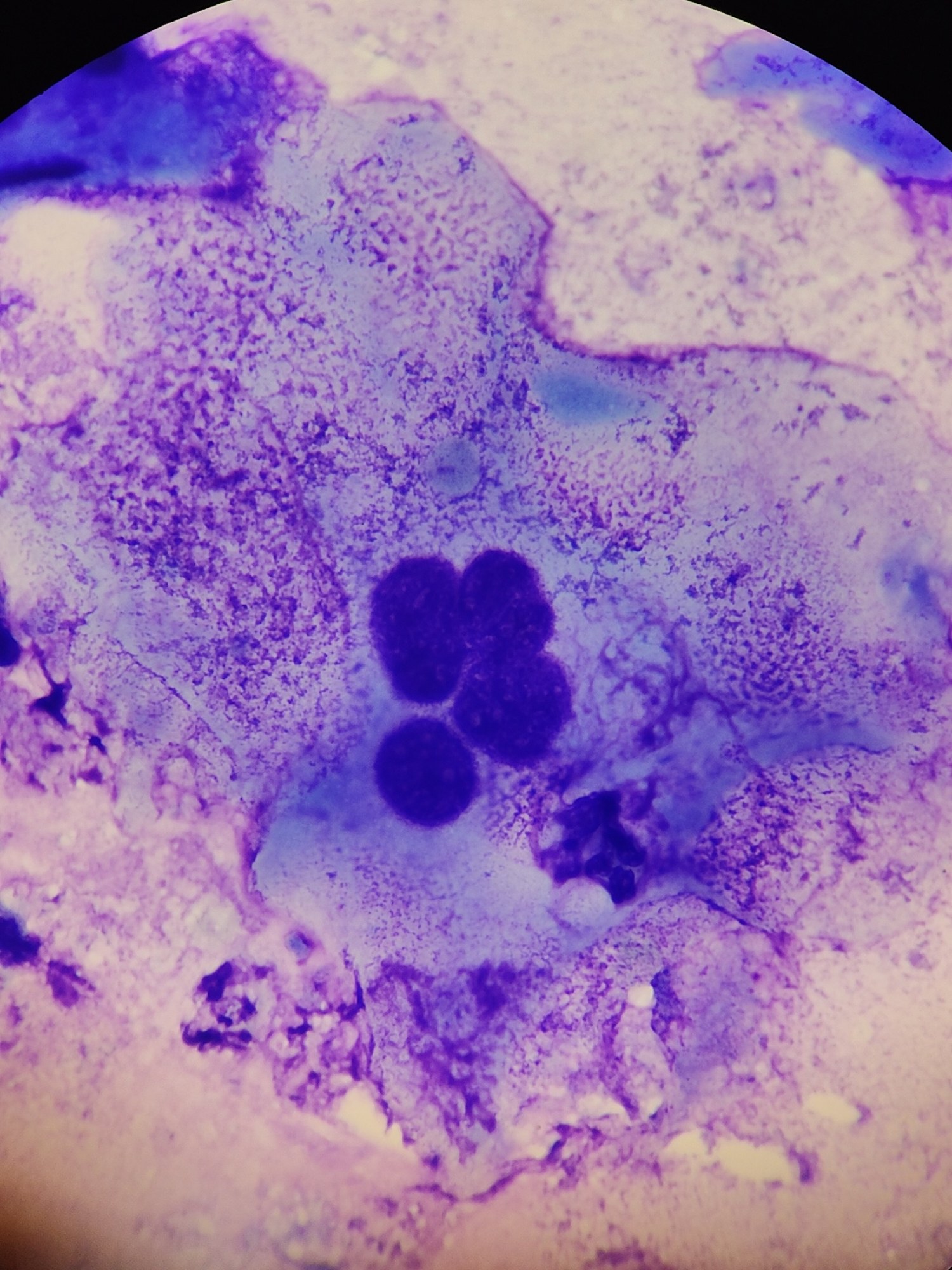
🔬 Cytology. Bronchoalveolar lavage. Confirmed squamous cell carcinoma with keratinization. Squamous cell carcinoma cells with keratinization are scattered throughout the preparation, in clusters, as microscopically small tissue fragments, and extremely rarely as “pearls” among sputum elements, mucus, and amorphous masses consisting of cytoplasmic remnants, scales, and cells with barely visible disappearing nuclei. Tumor cells vary in size (small, medium, and large) and shape (round, polygonal, elongated, ribbon-like, racket-shaped, etc.). Their nuclei are large, most often hyperchromatic, dense, with coarse, unevenly distributed chromatin, irregular contours, and highly diverse in shape (round, rod-shaped, triangular). Sometimes they occupy a small part of the cell. In cells undergoing necrobiosis, nuclei are destroyed and become barely visible. The cytoplasm in many cells is abundant, dense, shiny, often with signs of fatty degeneration.
🙏 Photos and description were kindly provided by biologist Anna Feditskaya, Luhansk.


