

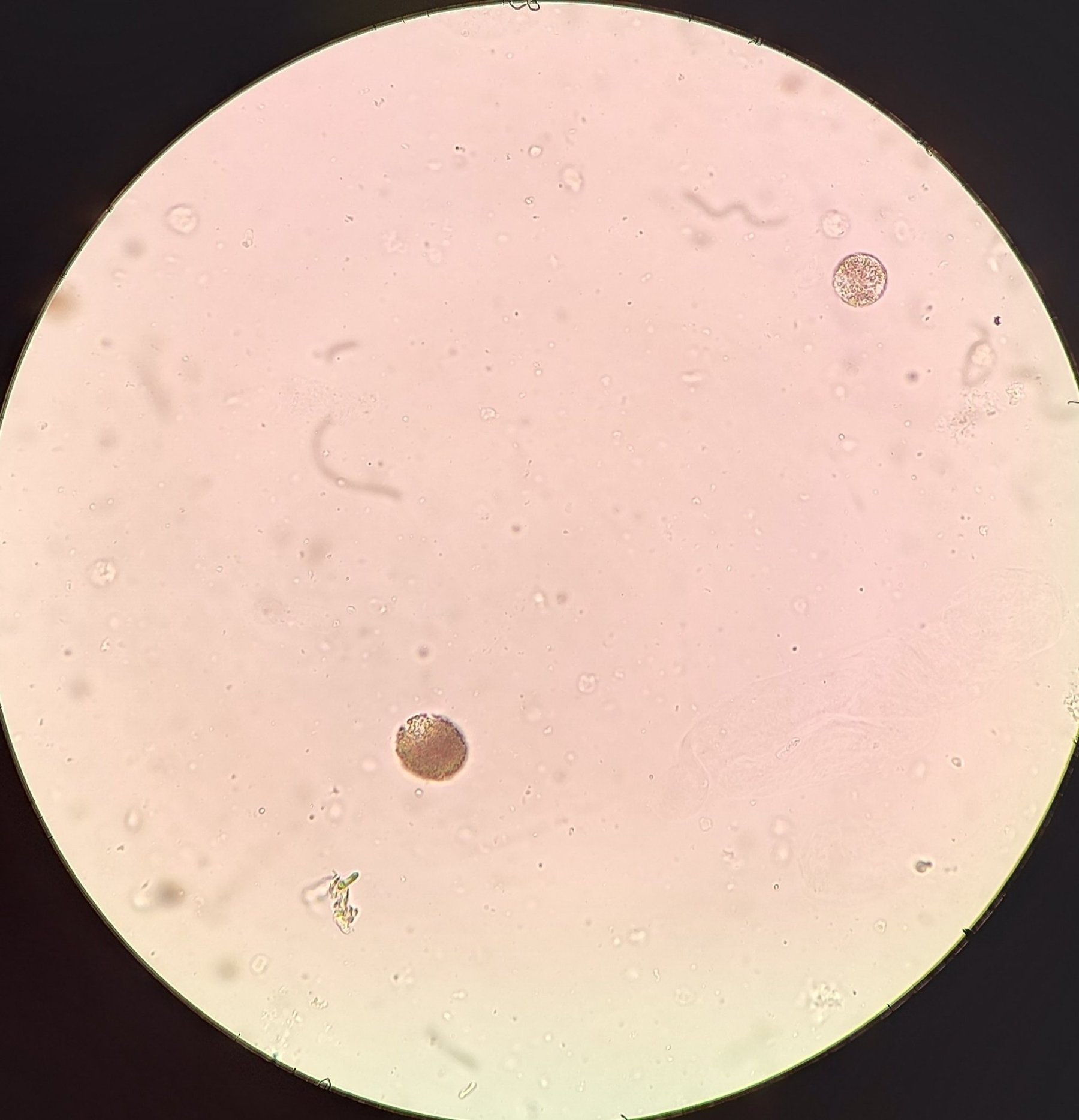
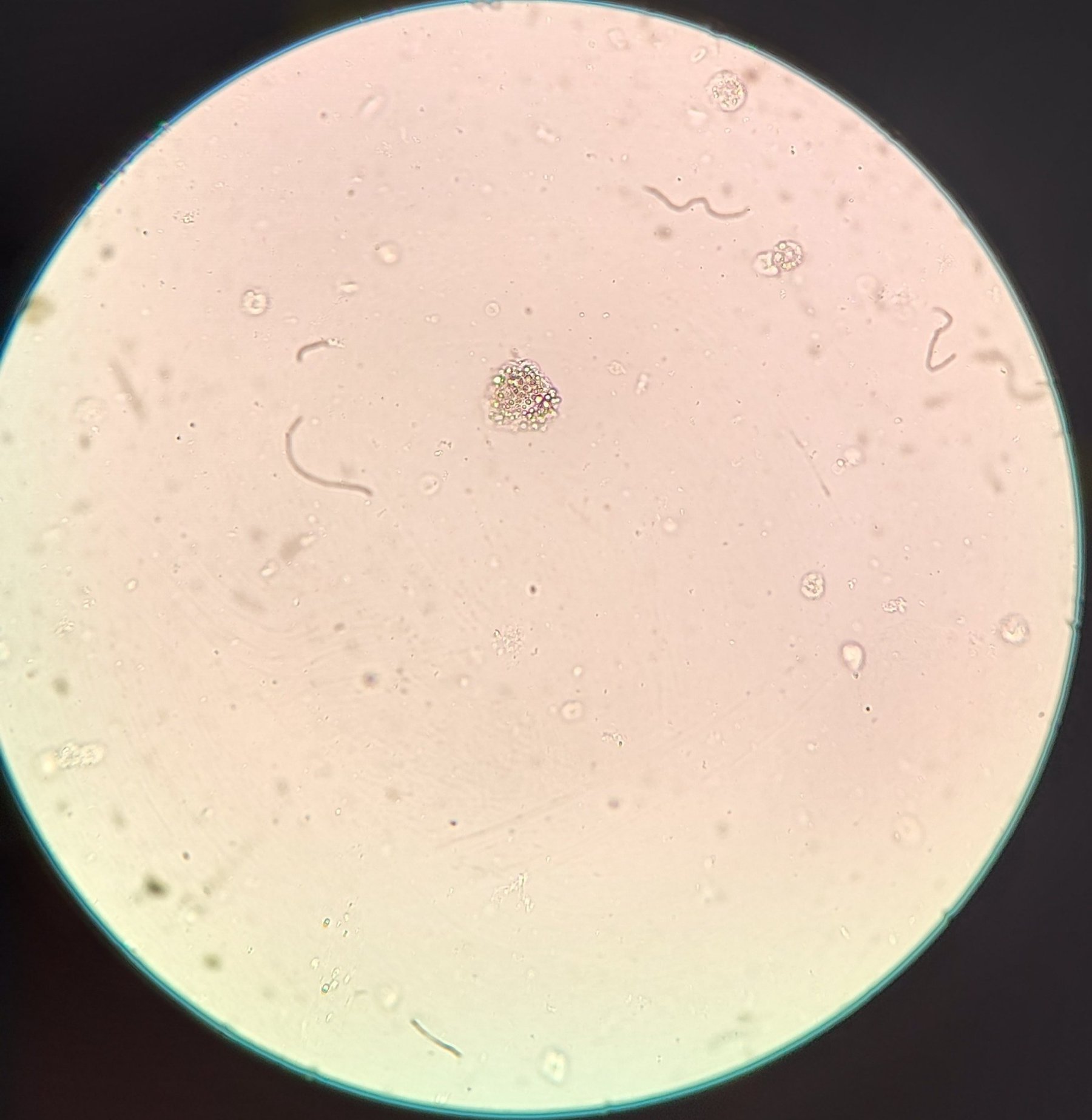
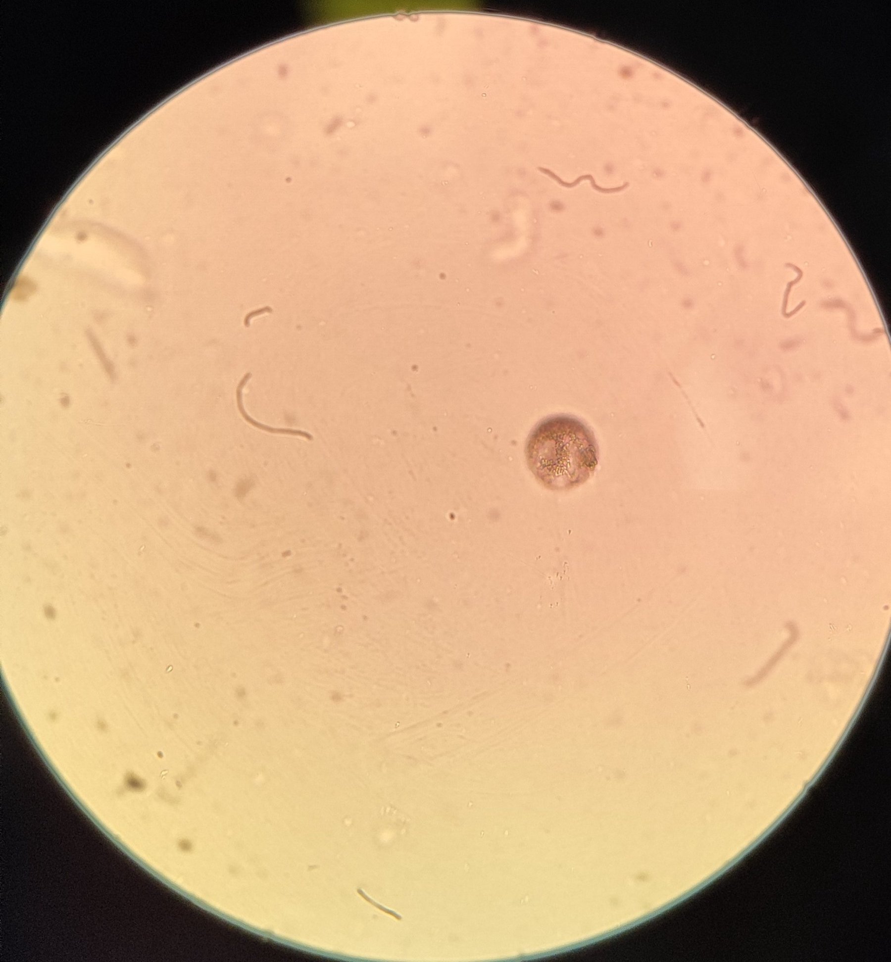
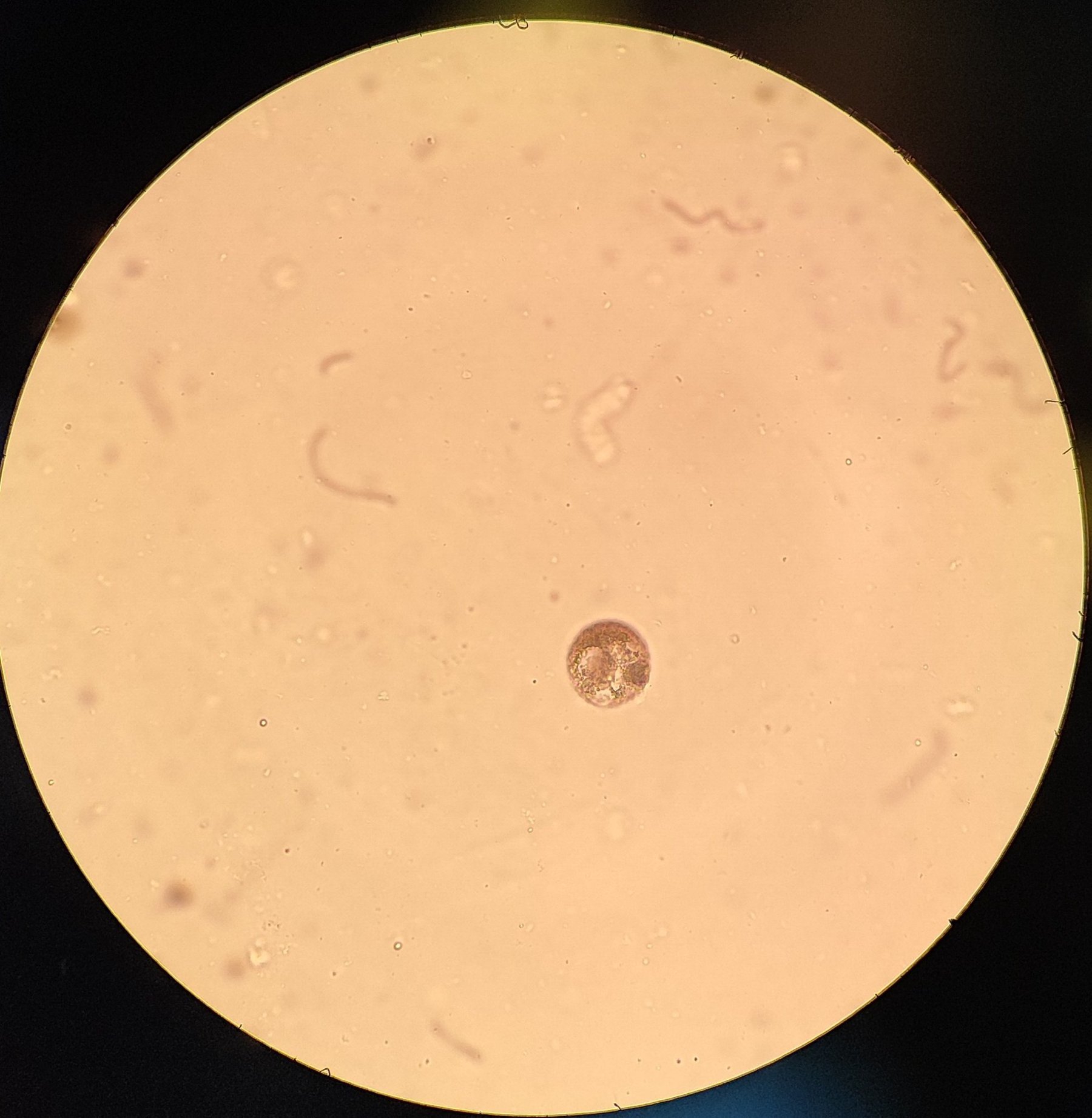
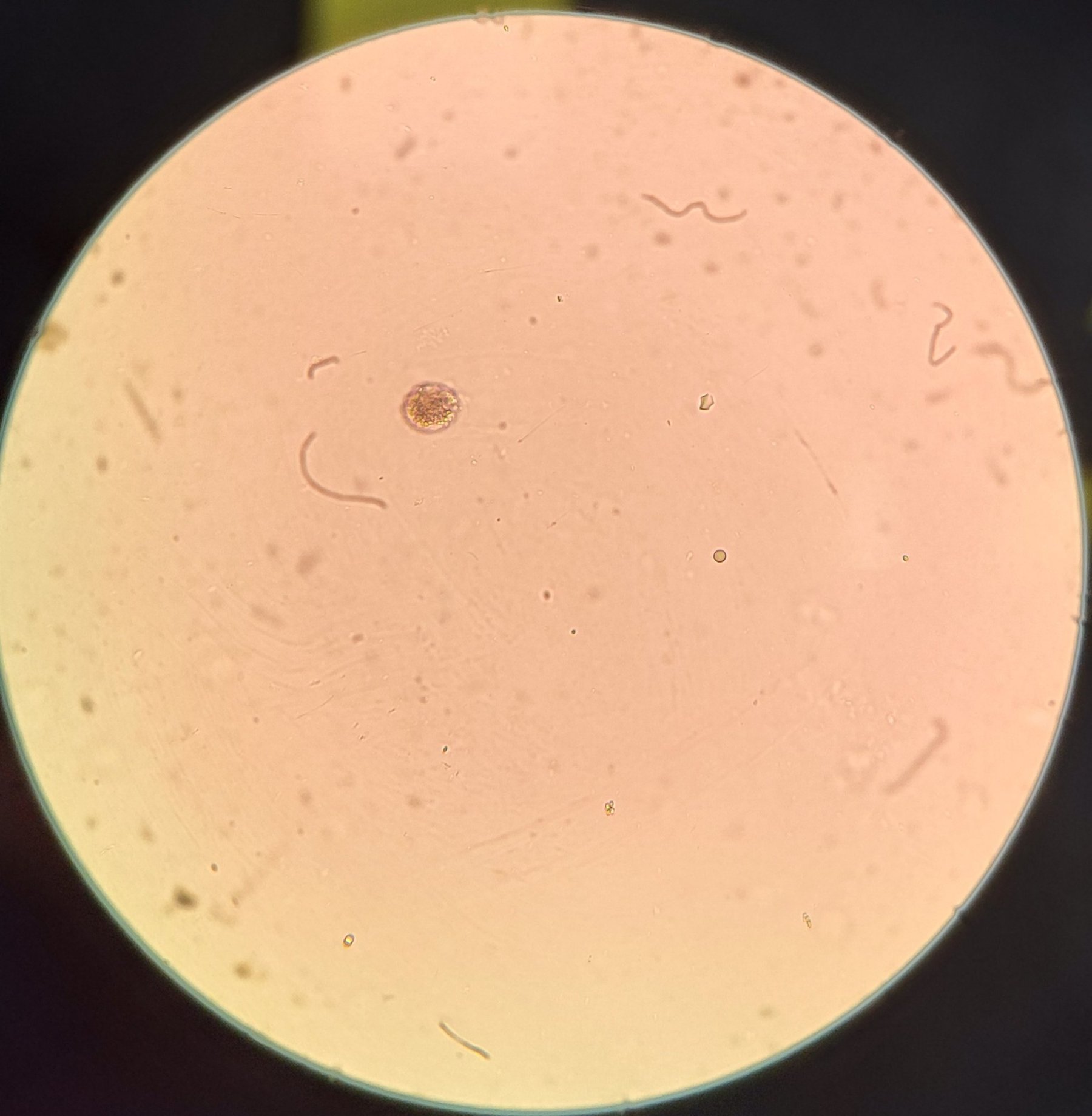
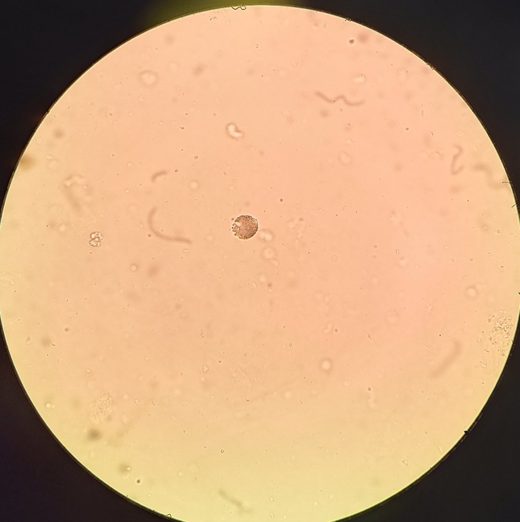
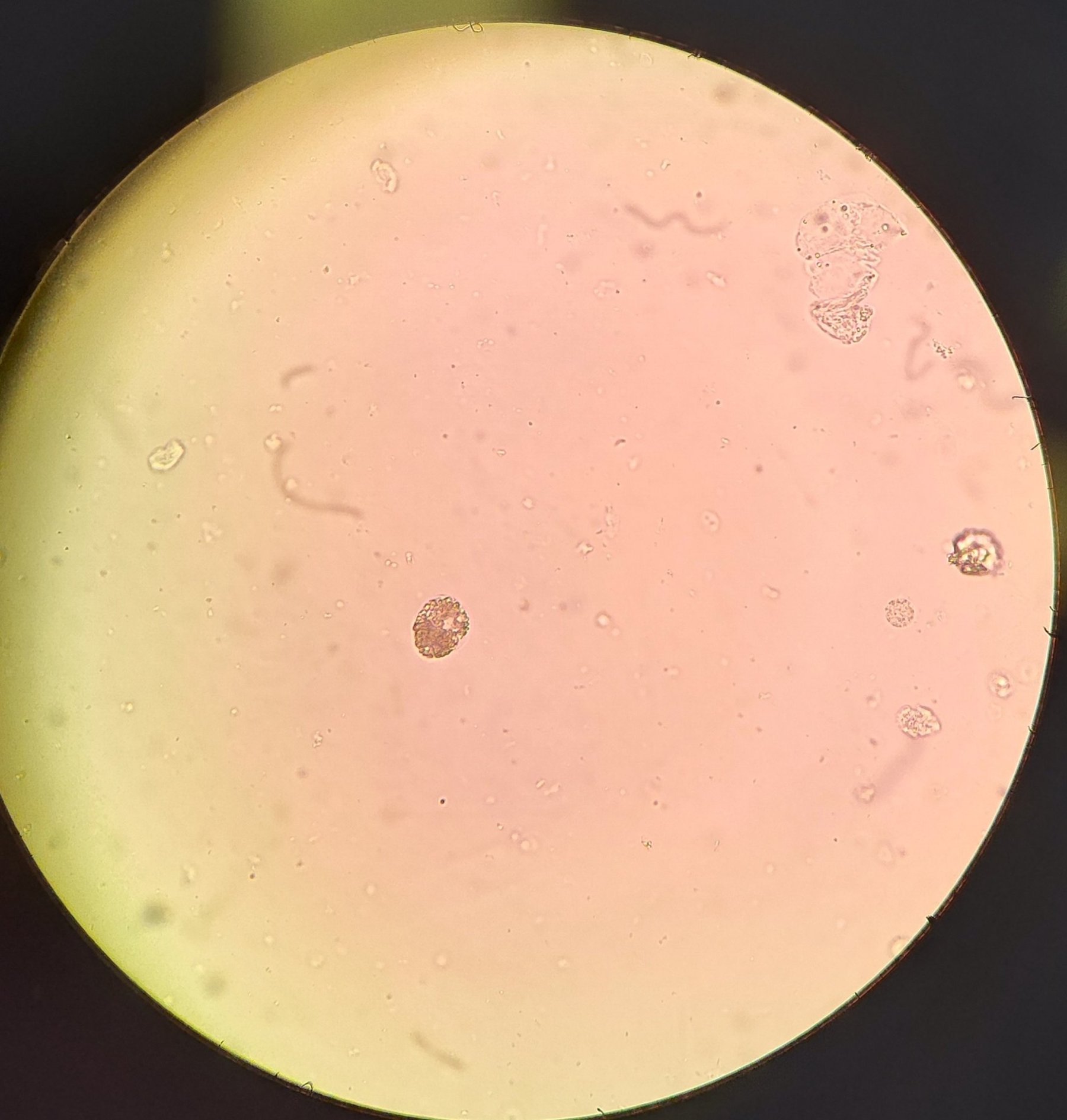
Renal epithelial cells with fatty degeneration. Cells are significantly enlarged (2-4 times larger than a leukocyte), densely packed with fat droplets. When viewed at low magnification, they strongly refracted light, appearing as dark brown spots. Native preparation, magnification x400
🙏 Material kindly provided by biochemist Daniil Adamov, Moscow


