

New article from a journal on cytopathology! 🔬 Polyomavirus in urine: when to sound the alarm? 🚨
A new study 🧐 has revealed important details about the diagnosis of polyomavirus (PV) in urine tests and its connection to bladder cancer. Here’s the main takeaway:
- 🦠 Type A (“classic PV”) — almost always benign, even if other suspicious cells are present.
- 🕸 Type B (“spiderweb chromatin”) — in 22% of cases, this may be cancer.
- ⚡ Type D (severely altered cells) — in 61.5% of cases, associated with a high risk of tumor.
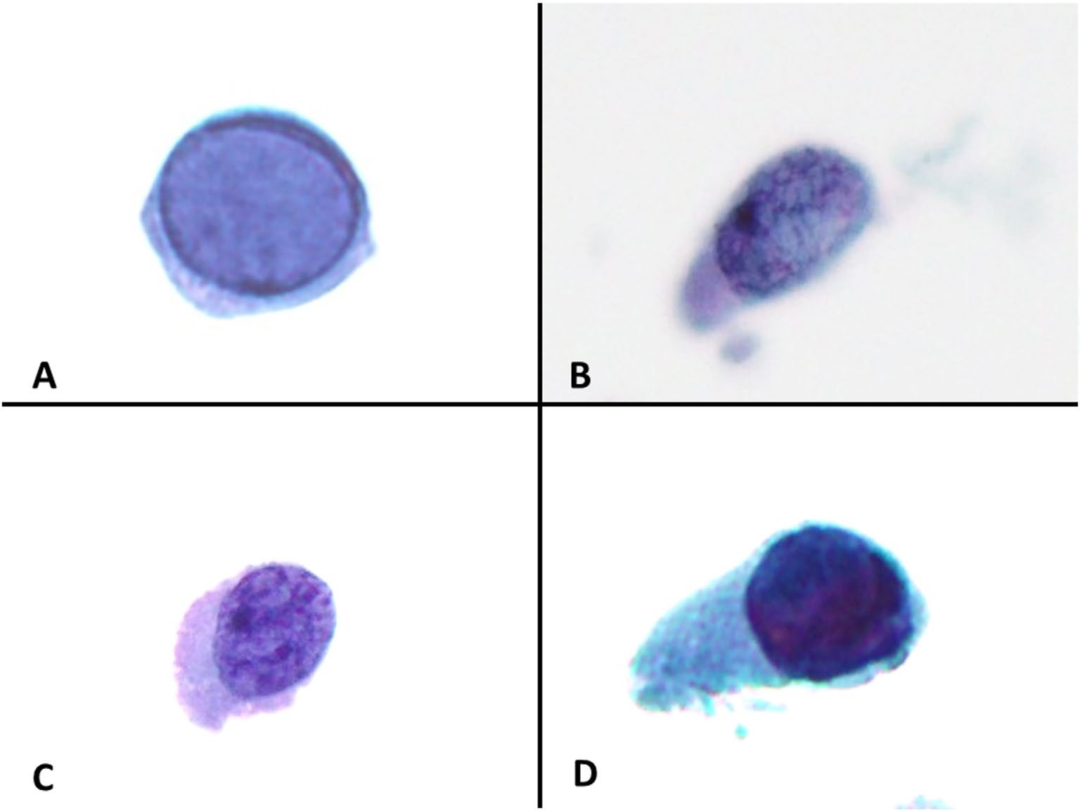
A. Type A cell — a single cell with changes typical of polyomavirus infection: a well-preserved cell with a blurred, homogeneous basophilic inclusion in the nucleus, with chromatin margination (displacement to the periphery) and smooth nuclear membranes.
B. Type B cell — a degeneratively altered cell with normo- or hyperchromatic nuclei, in which discernible chromatin details are preserved; the chromatin has a filamentous, “spiderweb” structure.
C. Type C cell — a degeneratively altered cell with normo- or hyperchromatic nuclei, in which chromatin details are also discernible; the chromatin has a granular or clumpy structure.
D. Type D cell — a degeneratively altered cell with homogeneous, sharply hyperchromatic nuclei, in which the chromatin pattern or distribution cannot be discerned.
Conclusions: 🔹 If type A cells are present in the analysis, the risk of cancer is extremely low (1.2%) — no need to worry. 🔹 Cells B, C, and D without type A — a reason for additional studies, especially in patients with a history of cancer.
📌 Source: Li Y. et al. Diagnostic Cytopathology, 2025.
@doc176287252_691104025 (Original article PDF→)


