

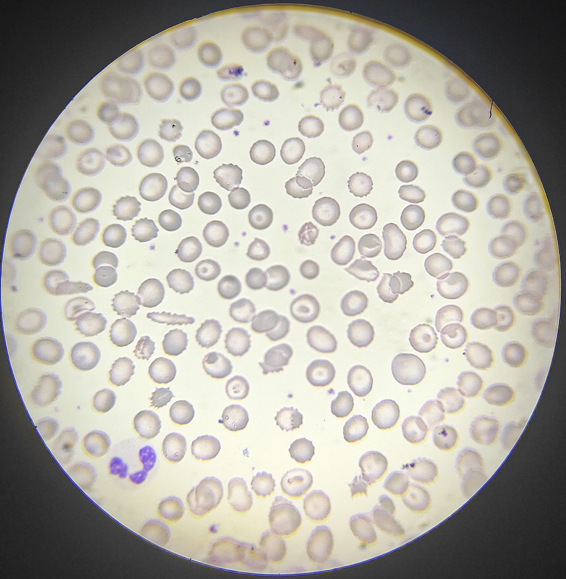
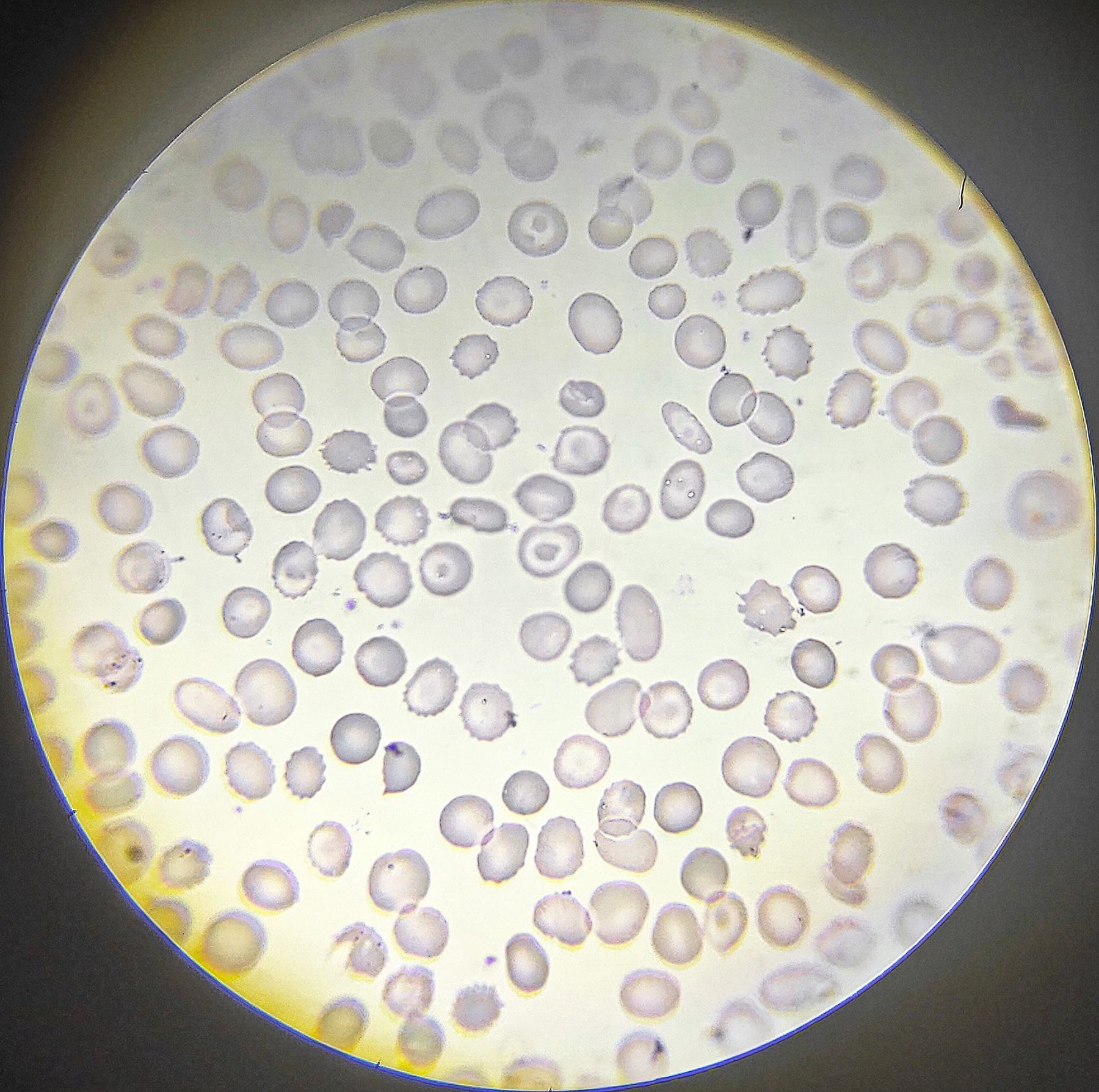
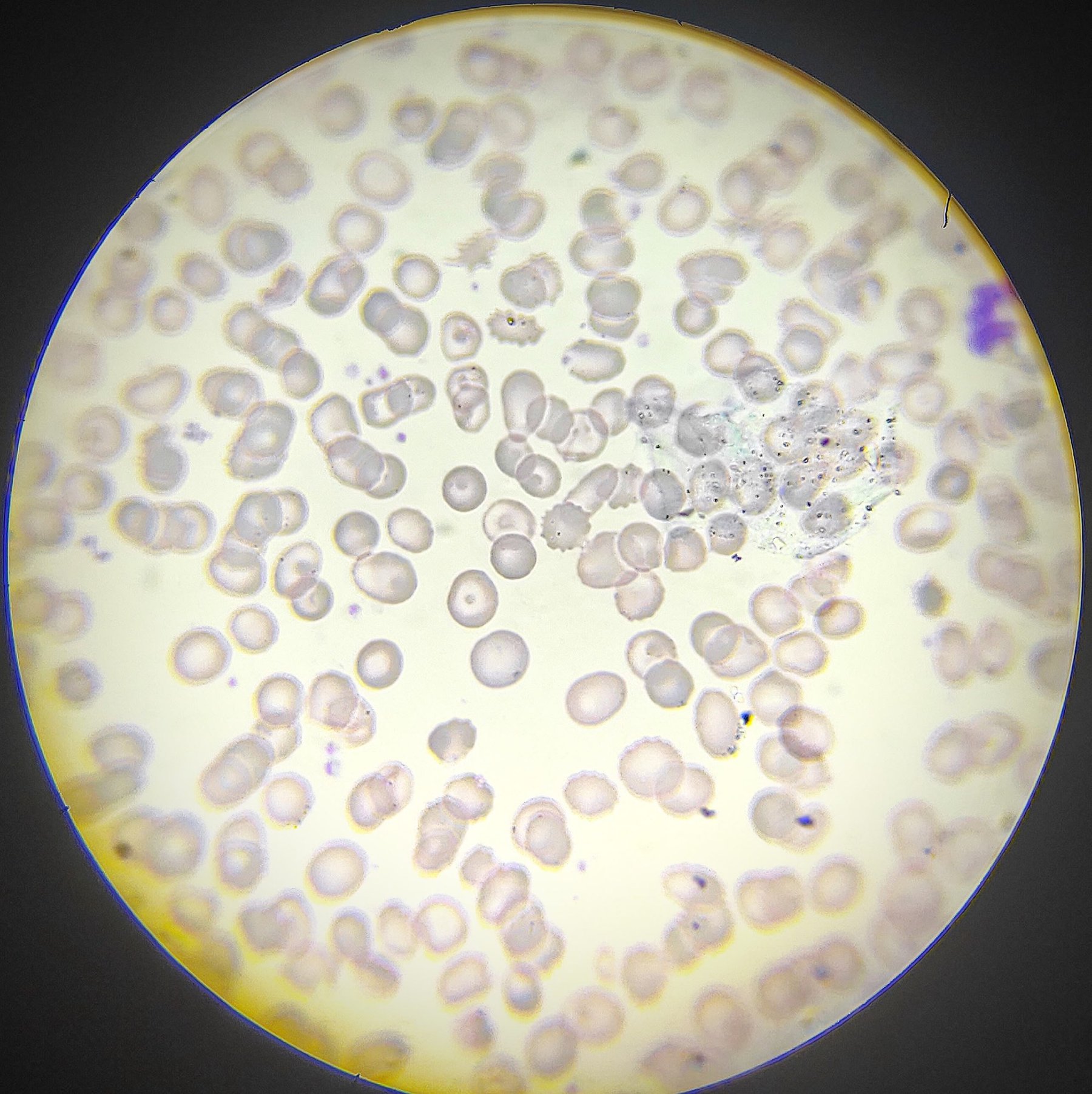
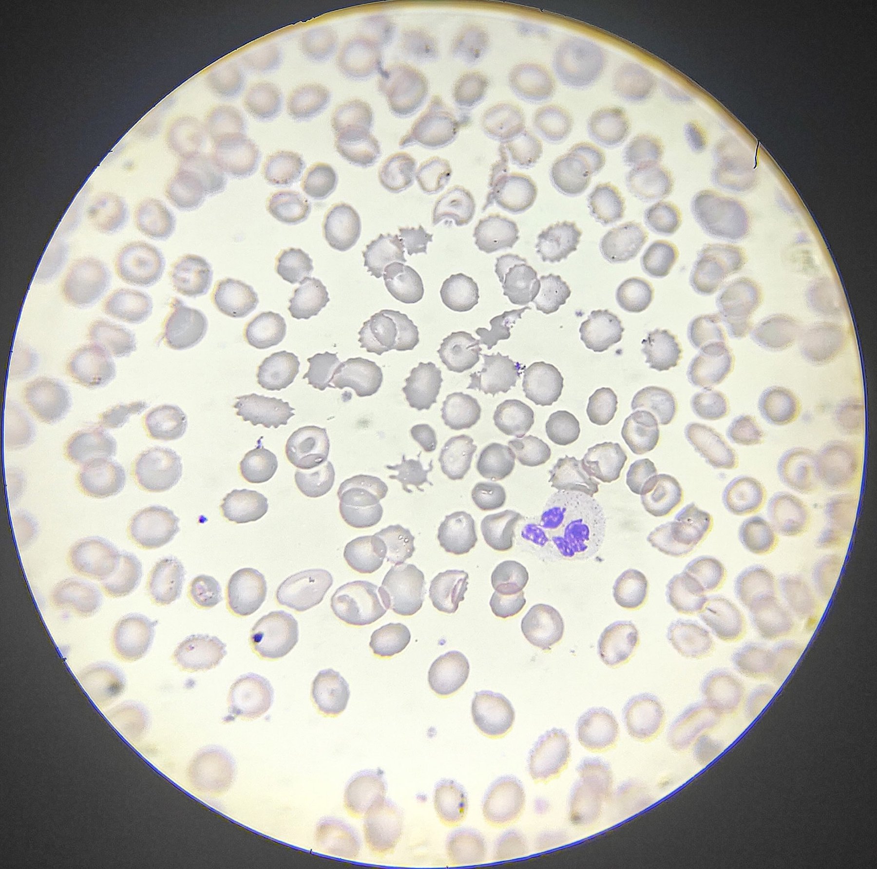
👴🏻Patient: 69 years old. History: gastrosplenectomy. 🩸In peripheral blood: POIKILOCYTOSIS.
🔹Codocytes (target cells) Erythrocytes with a stained center where hemoglobin is concentrated, giving them a visual resemblance to a 🎯.
The presence of these erythrocyte forms may indicate liver diseases, hemoglobinopathies, IDA, thalassemia, and splenectomy.
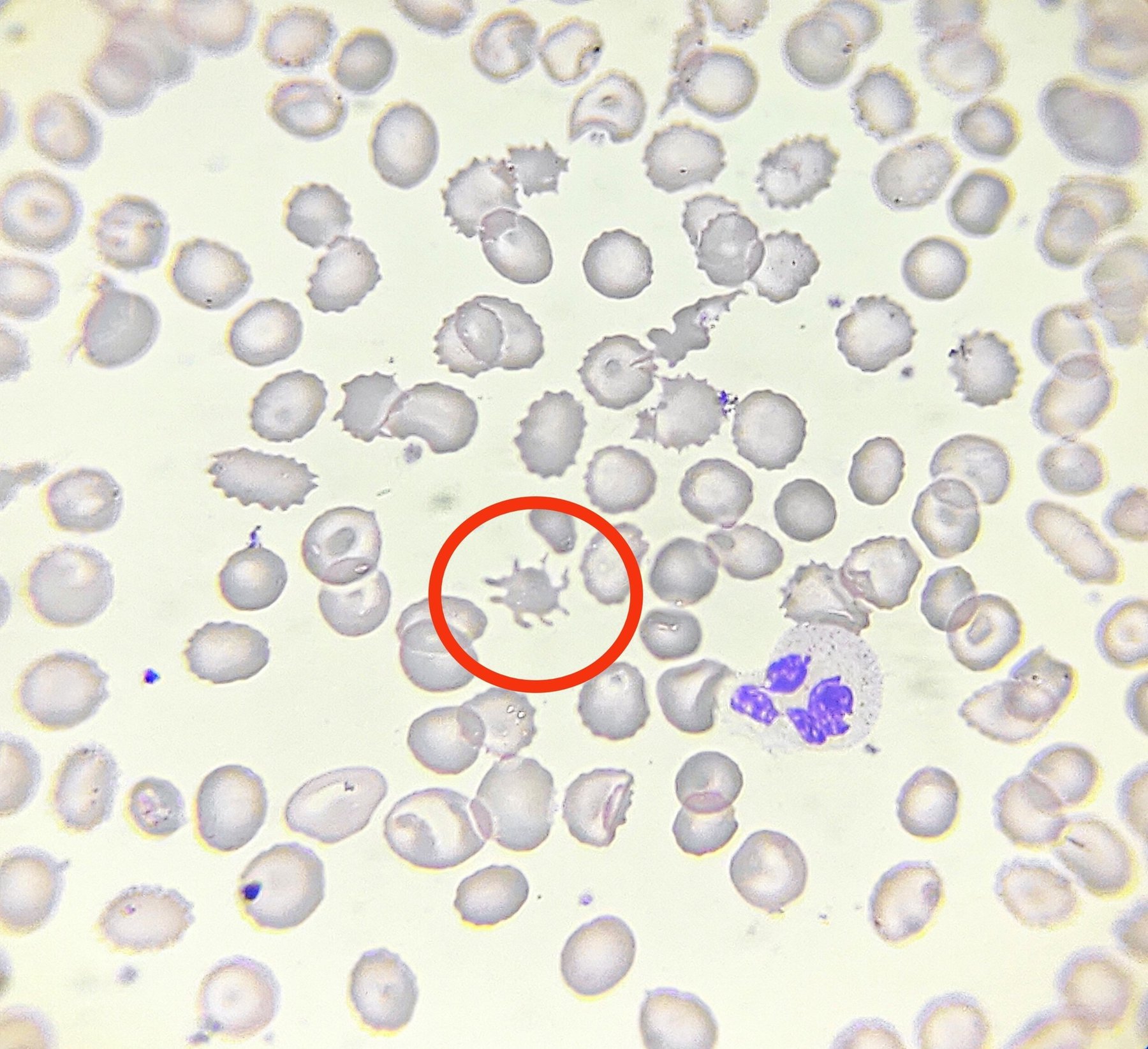
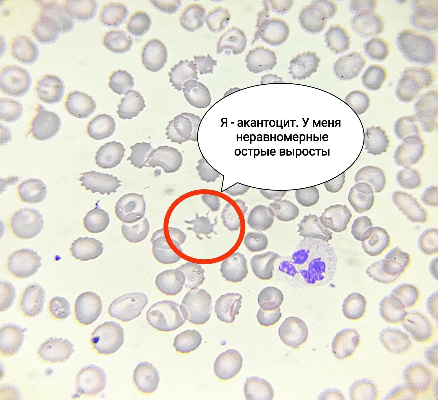
TASK from a subscriber! What is located in the very center of the smear in photos 4-5? Choose one answer:
- Spherocyte with outgrowths like a “jellyfish”
- Echinocyte
- Acanthocyte
- Schistocyte
☝🏻The answer is in photo 6.
🙏 The publication was kindly provided by clinical laboratory diagnostics physician Tanya Stepanova, Volgograd.
What other erythrocyte forms did you see?


