

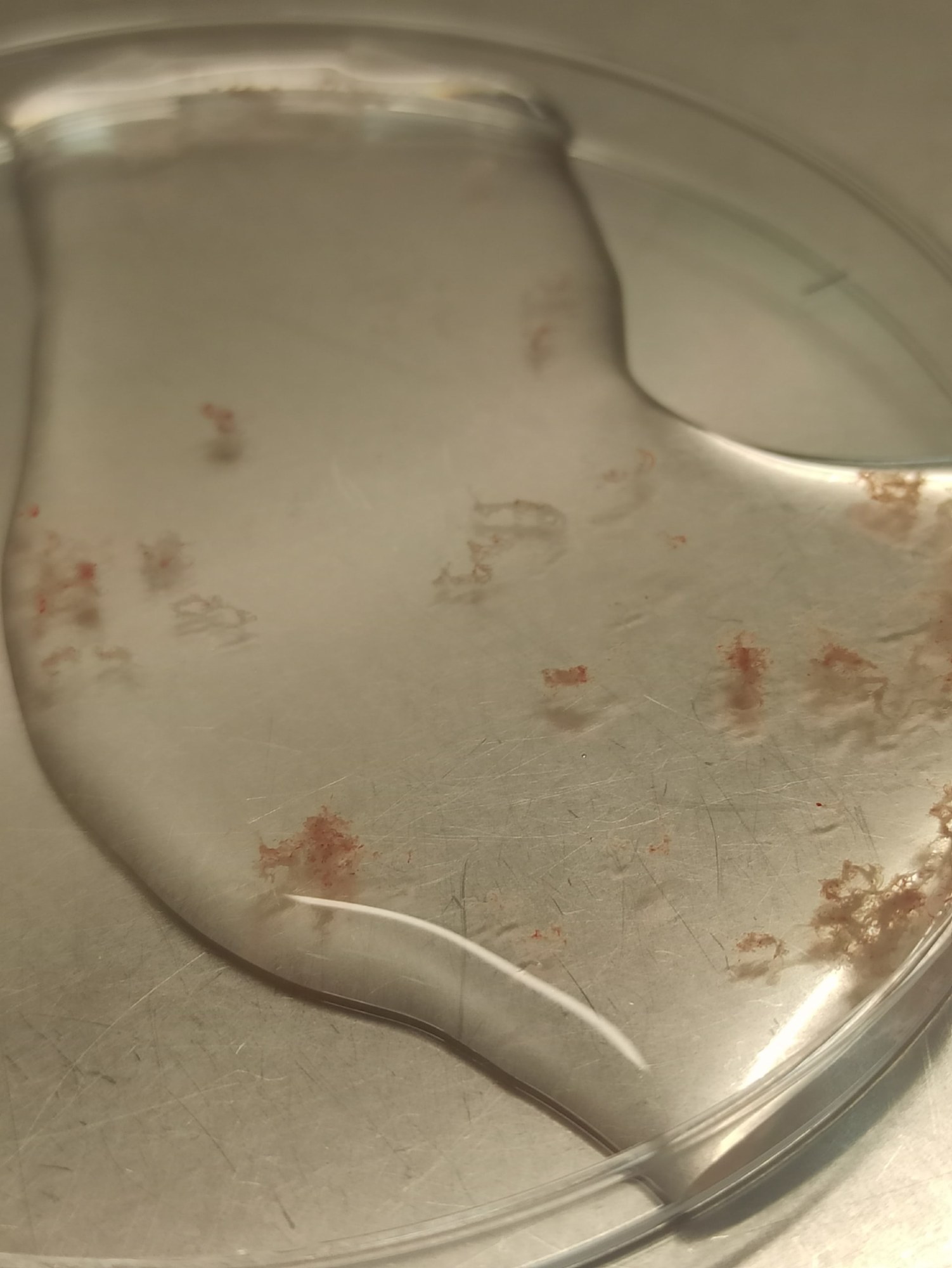
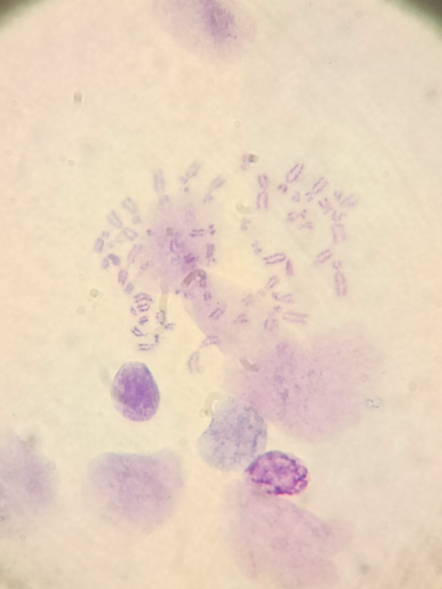
Accumulation of chromosomes in a metaphase plate (photo 2) in a karyotyping preparation made from chorionic villi (photo 1). Romanowsky staining. Chromosome visualization is achieved by treating the villi with various methods:
- Soaking in a hypotonic sodium citrate solution – promotes cell maceration (swelling).
- Colchicine treatment – destruction of spindle fibers, which stops cell mitosis at the metaphase stage.
- Mechanical cytolysis – after fixation, chromosomes are “shaken out” of the cells by gently tapping the villus against a glass slide in a drop of 60% acetic acid using forceps; the slide itself is preheated on an electric hot plate to 50 degrees Celsius.
- Proteolysis of intercellular connections and cell dissociation – achieved by treating the dried preparation with trypsin immediately before staining, thus separating the cells from each other, which is a necessary criterion for cytogenetic analysis. 🙏 Material kindly provided by medical laboratory technician Yelisey Zhigulyov, Ulyanovsk, perinatal center.


