

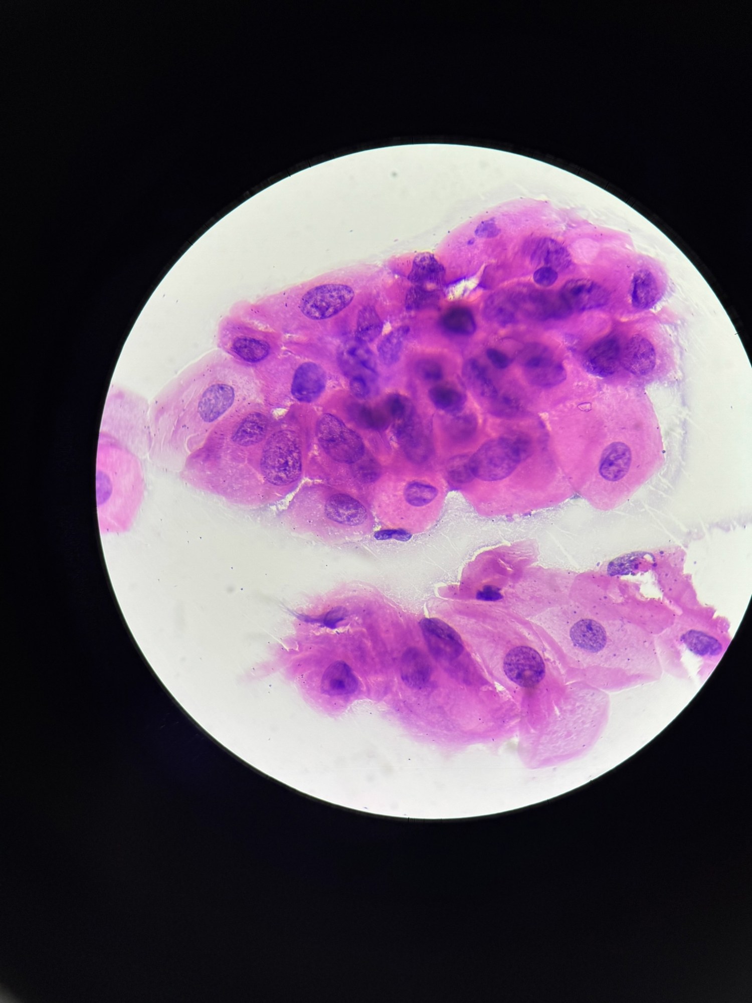
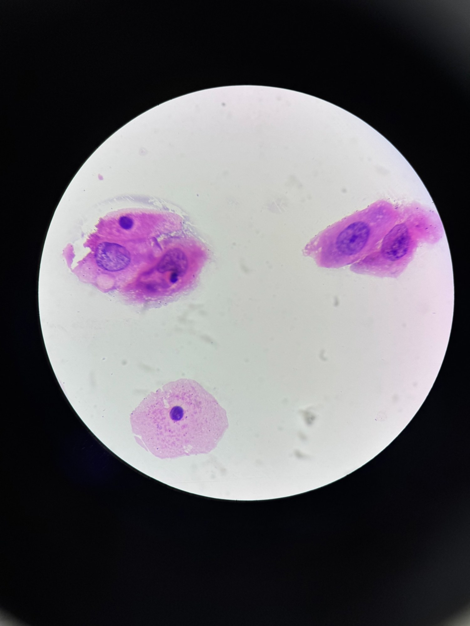
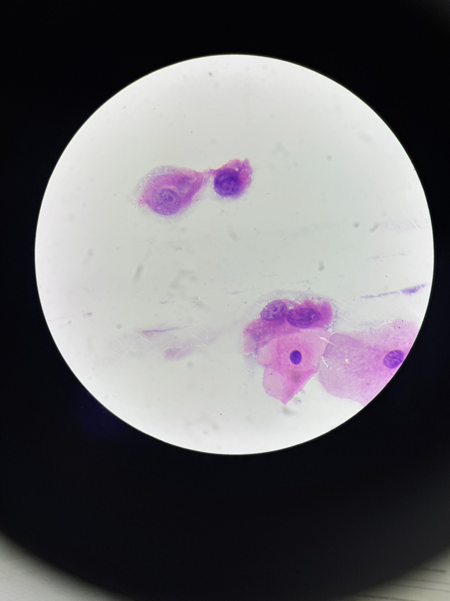
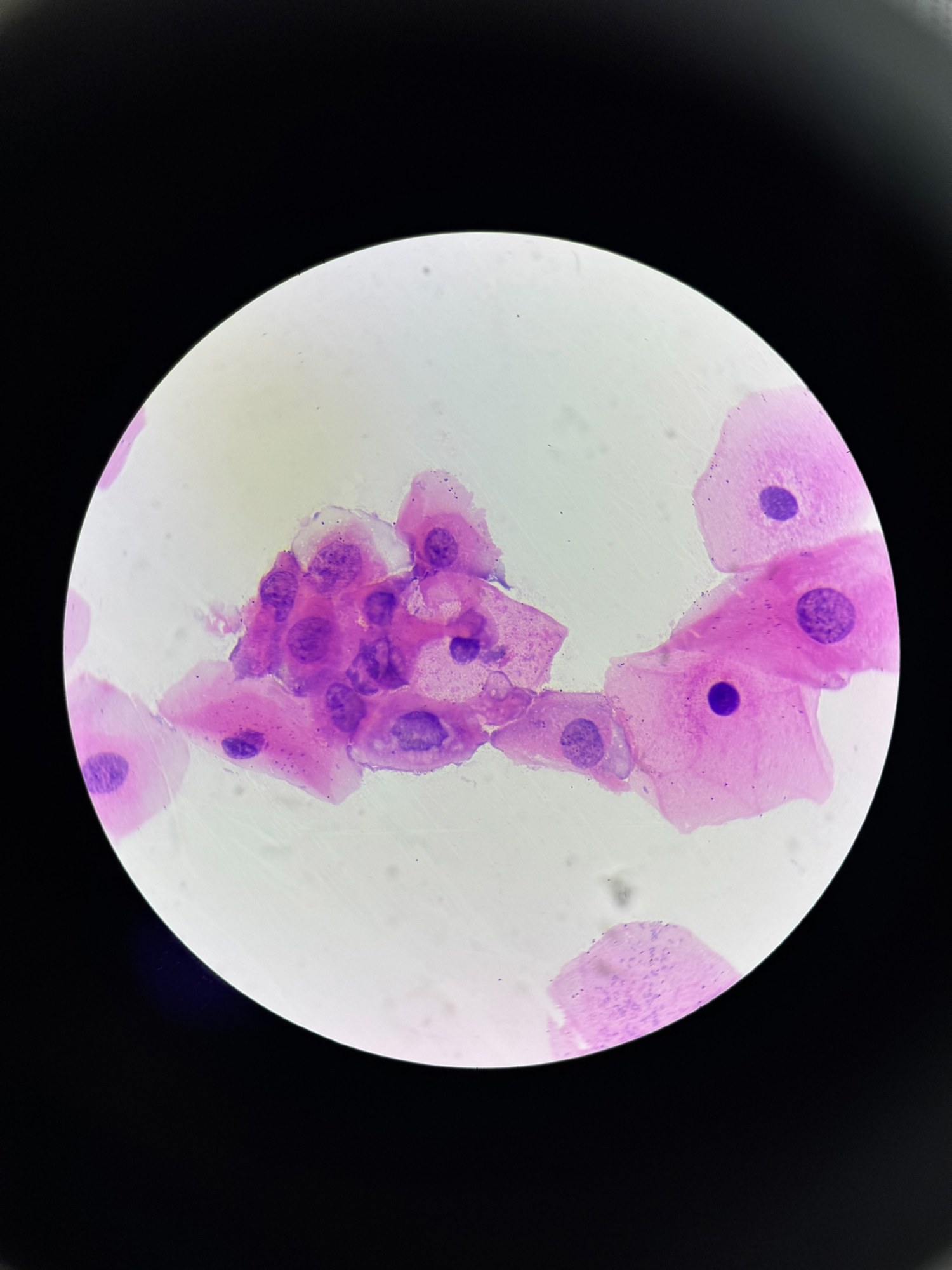
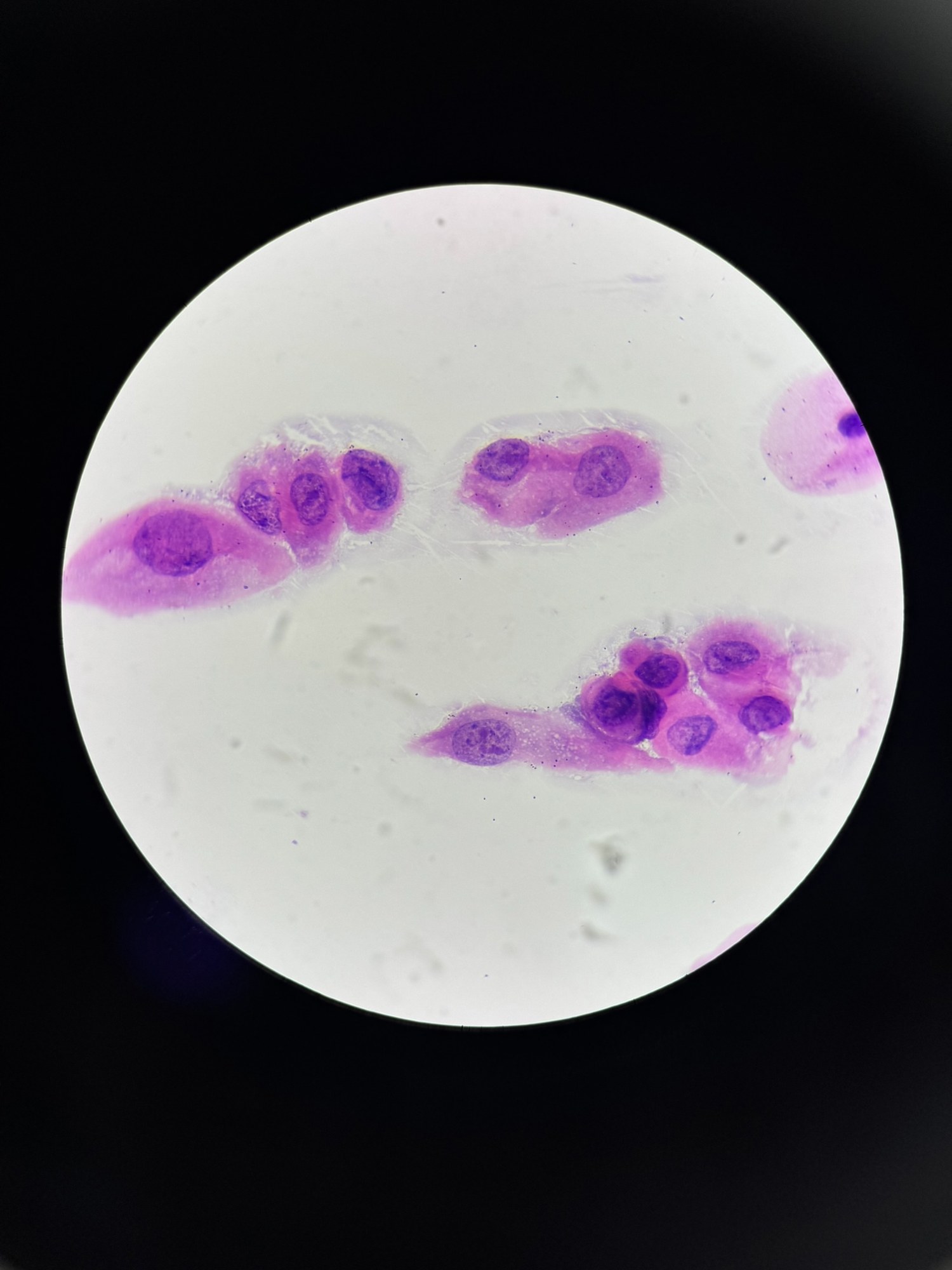
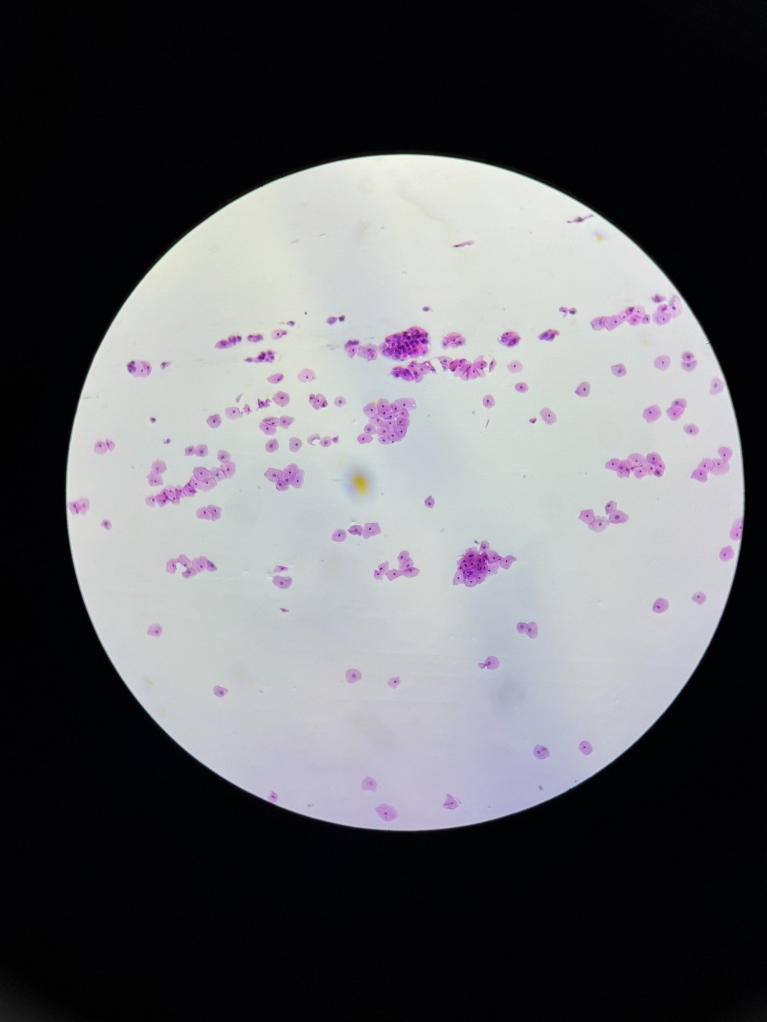
🔬 Cervical cytology. A woman of reproductive age, cytological examination during a preventive gynecological check-up, endocervix, LSIL
🙏 Smear photographs and description kindly provided by Kristina Yuryevna, clinical laboratory diagnostics physician, BU “RKB”, Cheboksary
💡 Background information: LSIL (Low-Grade Squamous Intraepithelial Lesion) is a term from a cytological report during cervical cancer screening. It indicates the presence of minor (low-grade) changes in epithelial cells, which may be caused by human papillomavirus (HPV).
📋 What does LSIL mean? LSIL is mild dysplasia or minimal changes in the squamous epithelium of the cervix. The main cause is infection with low- or high-risk oncogenic HPV. It is common in women of reproductive age. These changes can spontaneously regress (in 60-80% of cases), but require observation. 🔬 Cytological features of LSIL Atypical squamous epithelial cells with signs of koilocytosis (enlarged nucleus, peripheral halo). Cytoplasm remains relatively preserved. Changes in cells are often associated with virus replication in the epithelium. 🔍 Management of LSIL Follow-up cytological examination in 6-12 months. If high-risk oncogenic HPV is detected, colposcopy may be indicated to clarify the diagnosis. In case of LSIL persistence for more than 2 years – biopsy to rule out progression to HSIL. 🧬 Risk of progression LSIL rarely progresses to cancer, but in 10-20% of cases, it can progress to more severe changes (HSIL or CIN II/III). Regular observation and adherence to doctor’s recommendations are extremely important. 💡 Advice for patients: LSIL is not a verdict, but merely a signal for careful observation and health care. In most cases, timely monitoring and immune support help the body cope with the infection on its own.


