

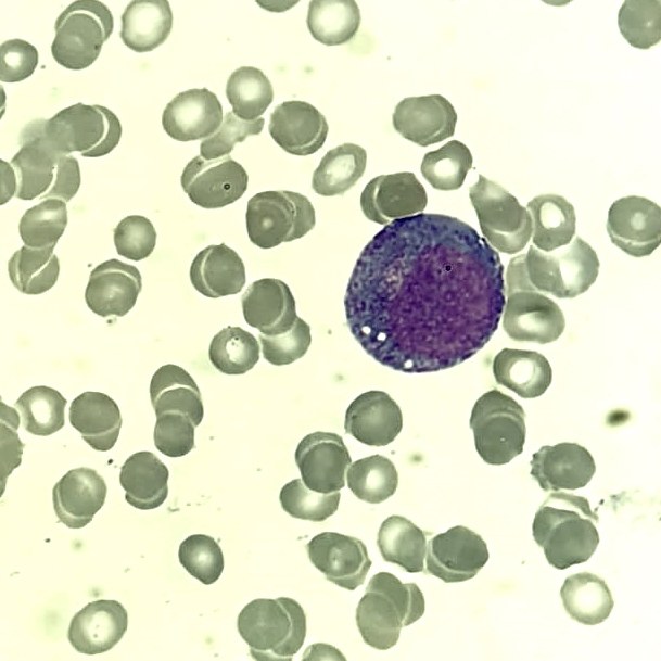
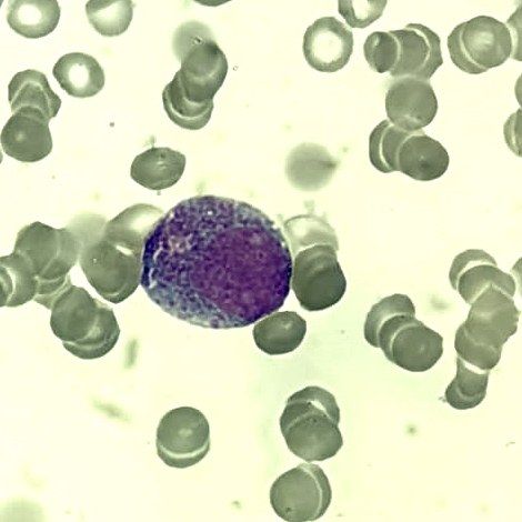
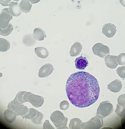
🔬 Classic promyelocytes. Microscopy of bone marrow aspirate from a patient with agranulocytosis (severe neutropenia).
✅ The cytoplasm is abundantly filled with large azurophilic granules, the nucleus has evenly dispersed chromatin, and there is a clearly defined perinuclear clearing zone.
✅ In autoimmune neutropenia, the bone marrow typically maintains a normal or increased number of granulocytic precursor cells. This is because the problem is not related to impaired production but to increased peripheral destruction of neutrophils under the influence of autoantibodies.
Characteristic changes in the bone marrow in autoimmune neutropenia:
👉 Granulocytic hyperplasia: Increased number of immature neutrophil forms (myeloblasts, promyelocytes, myelocytes). In some cases, an increased content of mature neutrophils may be observed, compensatory for peripheral loss. 👉 Absence of signs of aplasia or hypoplasia: Unlike aplastic anemia or toxic bone marrow damage, megakaryocytic and erythroid lineages remain preserved. 👉 Maturation features: Mature neutrophils may be morphologically normal, but their number in peripheral blood is reduced due to autoimmune destruction. 👉 Signs of regenerative stress: Sometimes, accelerated maturation of granulocytic cells is noted.
Thus, in autoimmune neutropenia, the main focus is on peripheral blood, while the bone marrow remains functionally active.
🙏 Material kindly provided by clinical laboratory physician Andrey Rudinsky, Hadassah Medical Moscow.


