

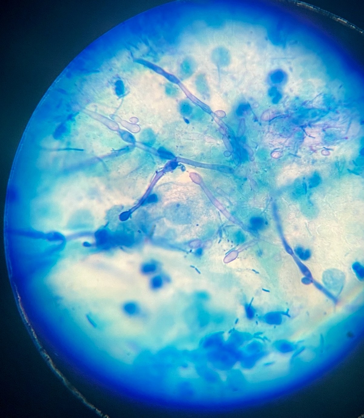
“Flourishing” yeast fungus in a vaginal smear.
🙏 The image was kindly provided by Clinical Laboratory Diagnostics physician Alena Chirvina, Rostov-on-Don.
Microscopic image of candidiasis in a vaginal smear: detection of Candida pseudomycelium and budding cells during flora smear microscopy.
“Flourishing” yeast fungus in a vaginal smear.
🙏 The image was kindly provided by Clinical Laboratory Diagnostics physician Alena Chirvina, Rostov-on-Don.

Microscopic view of candidiasis: detection of pseudomycelial filaments and Candida yeast-like fungal spores in a vaginal flora smear.

Morphology of Candida yeast-like fungi in a stained smear: detection of pseudomycelial filaments and budding blastospores by light microscopy.

Microscopic view of candidiasis: abundant accumulation of Candida pseudomycelia and blastospores in a gynecological vaginal smear. Signs of severe mycosis.