

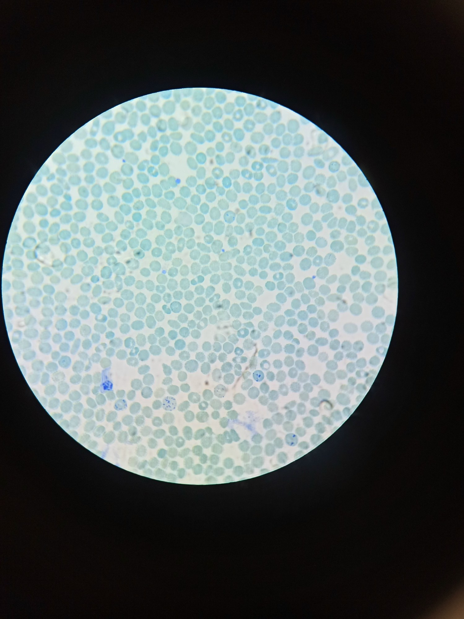
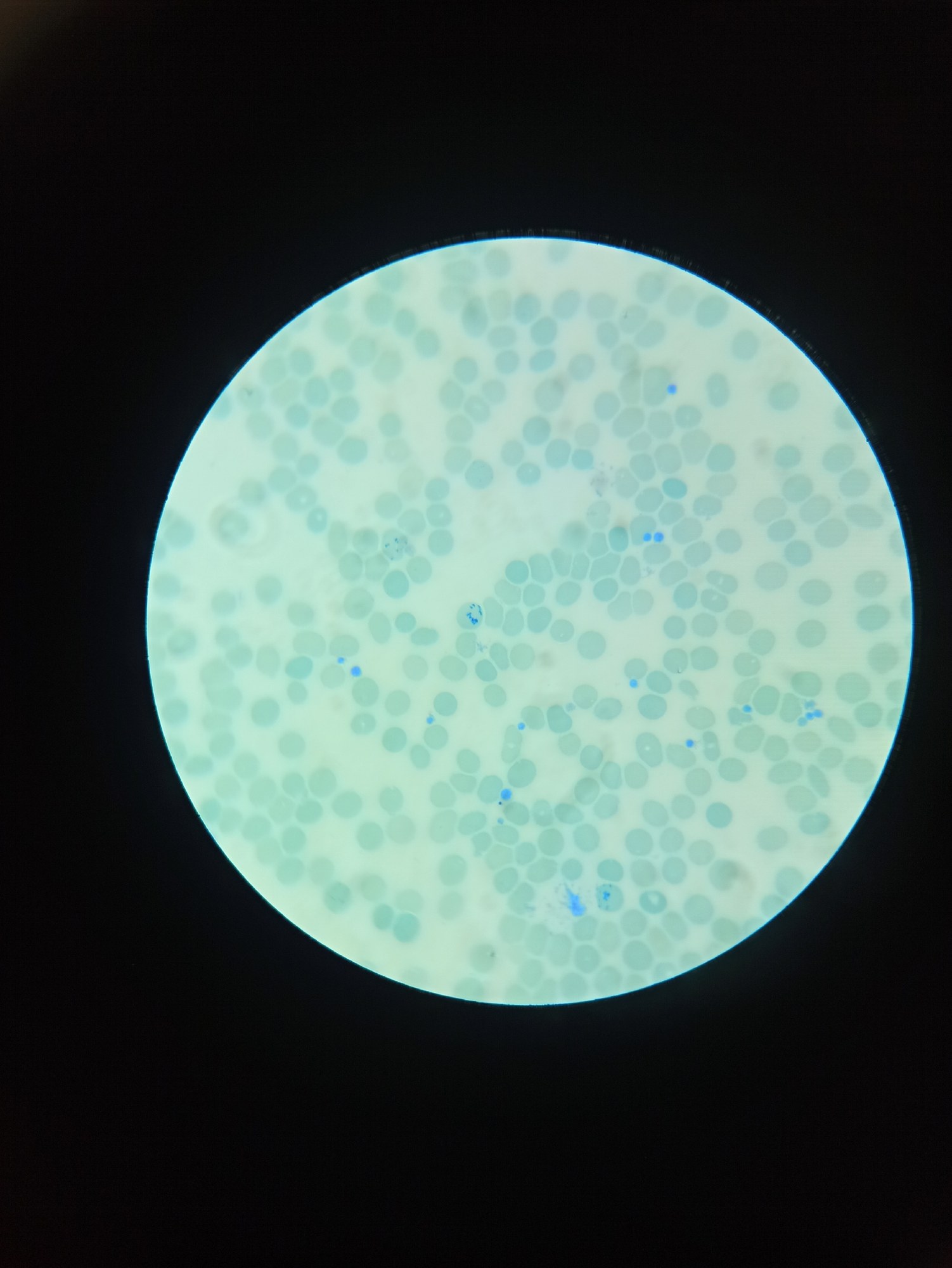
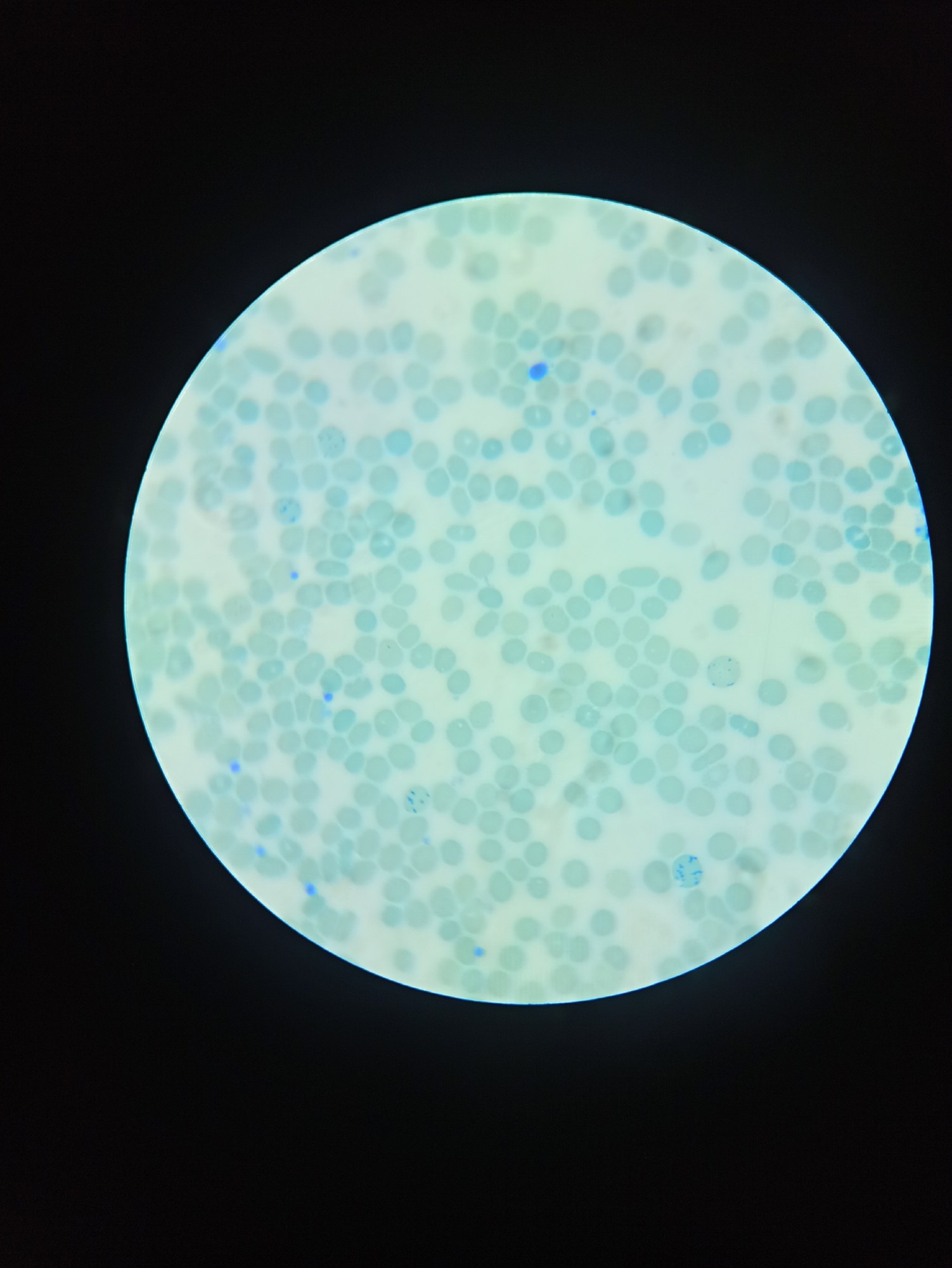
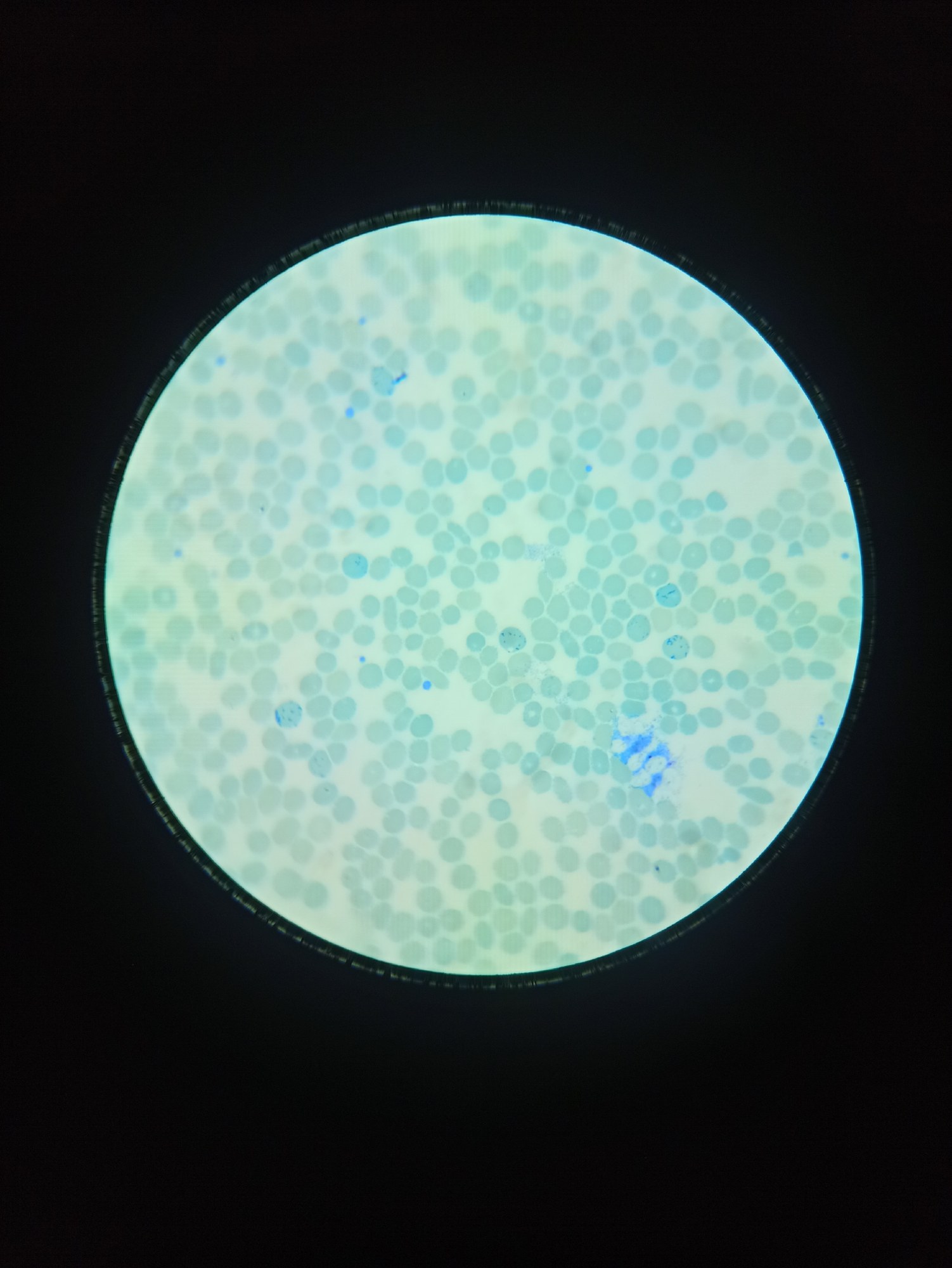
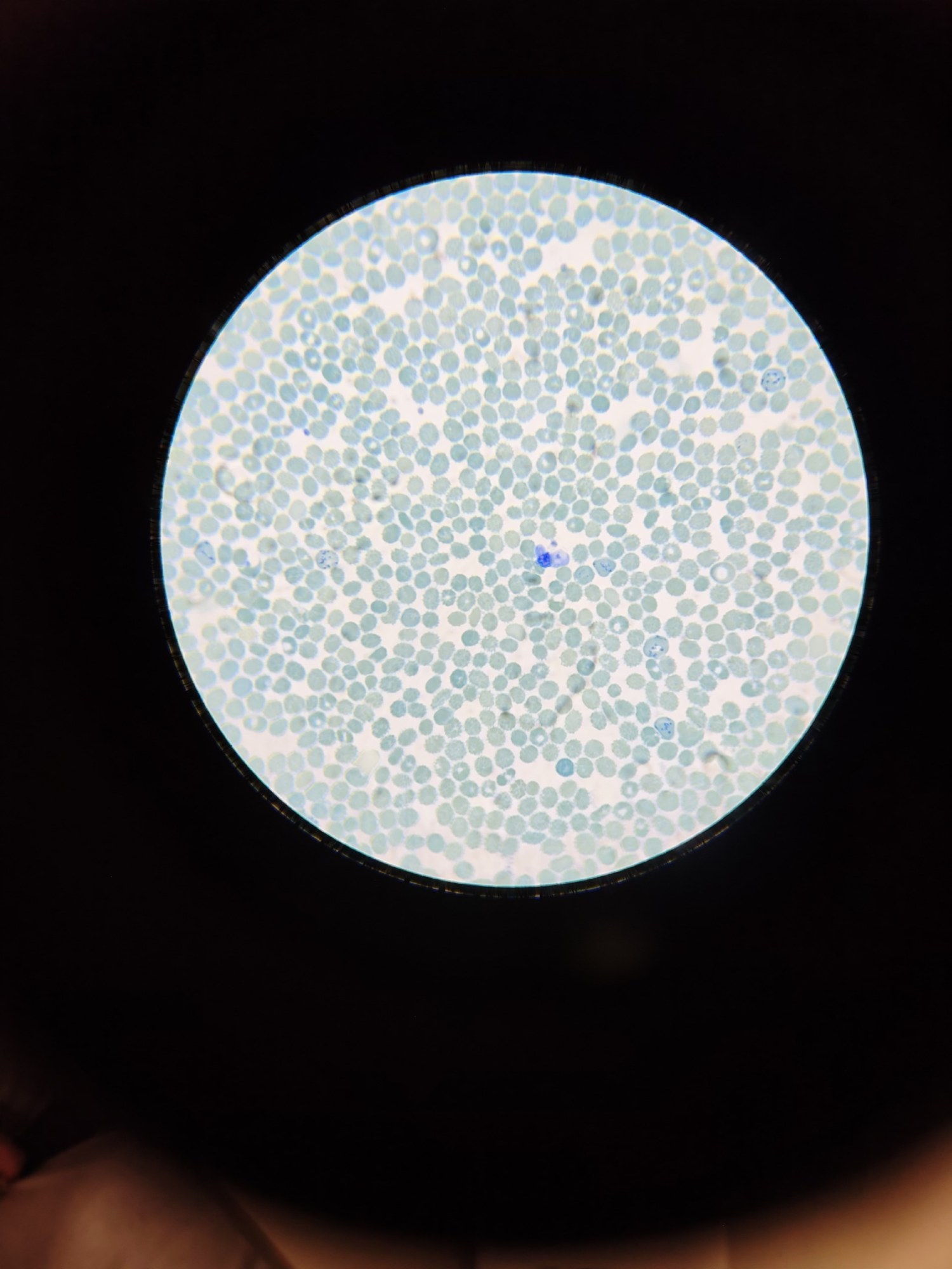
Reticulocytes, peripheral blood, brilliant cresyl blue stain.
Reference information. Reticulocytes are immature cells of the erythroid (red) hematopoietic lineage; they occupy an intermediate position between the oxyphilic erythroblast (normoblast) and the mature erythrocyte.
After the spleen removes the nucleus, the reticulocyte retains ribosomal remnants for hemoglobin synthesis, while a mature erythrocyte cannot synthesize hemoglobin independently, but only transports existing hemoglobin.
Detection methods. Most often, supravital (live – without additional fixation) staining with methylene blue or brilliant cresyl blue is used to detect reticulocytes. With this method, blue structures – ribosomal remnants – can be found inside reticulocytes. However, reticulocytes can also be detected with conventional staining: since ribosomes are still present in these cells and protein biosynthesis continues, reticulocytes retain an affinity for basic dyes such as azure 1, azure 2, or methylene blue. Thanks to this, when stained by the Romanowsky or May-Grünwald method, reticulocytes appear as erythrocytes that are slightly larger (approximately 9-11µm versus 7-8µm for a normal erythrocyte) and have darker cytoplasm with a lilac tint (so-called polychromatophilic erythrocytes), whereas in mature erythrocytes, protein biosynthesis is completely ceased, which causes them to lose affinity for basic dyes and only absorb acidic dyes, such as eosin.
Diagnostic significance. Normally, the number of reticulocytes circulating in peripheral blood is 2-10 per mille (0.2-1%). Physiological reticulocytosis is observed during treatment of anemias with iron preparations, vitamin B9 (folic acid), B12 (cyanocobalamin), after erythropoietin administration, in young children, during pregnancy, and in conditions of tissue hypoxia.
Pathological reticulocytosis is characteristic of acute blood loss, true polycythemia (erythremia), as well as conditions and diseases with increased hemolysis – hemolytic anemias, thrombotic thrombocytopenic purpura (TTP), hemolytic uremic syndrome (HUS), paroxysmal nocturnal hemoglobinuria (PNH), march hemoglobinuria, crush syndrome (CS), hemolytic disease of the newborn (HDN), transfusion of ABO/Rh incompatible blood.
Reticulocytopenia is characteristic of pathologies with erythroid lineage suppression or ineffective erythropoiesis, such as kidney diseases (suppression of erythropoietin synthesis), hemophagocytic lymphohistiocytosis (HLH), aplastic anemia, megaloblastic anemia, parvovirus B19 infection, malignant neoplasms, including bone marrow metastases (with the exception of true polycythemia), radiation sickness.
🙏 The material was kindly provided by medical laboratory technician of Neva Clinic Elisey Zhigulyov, Ulyanovsk


