

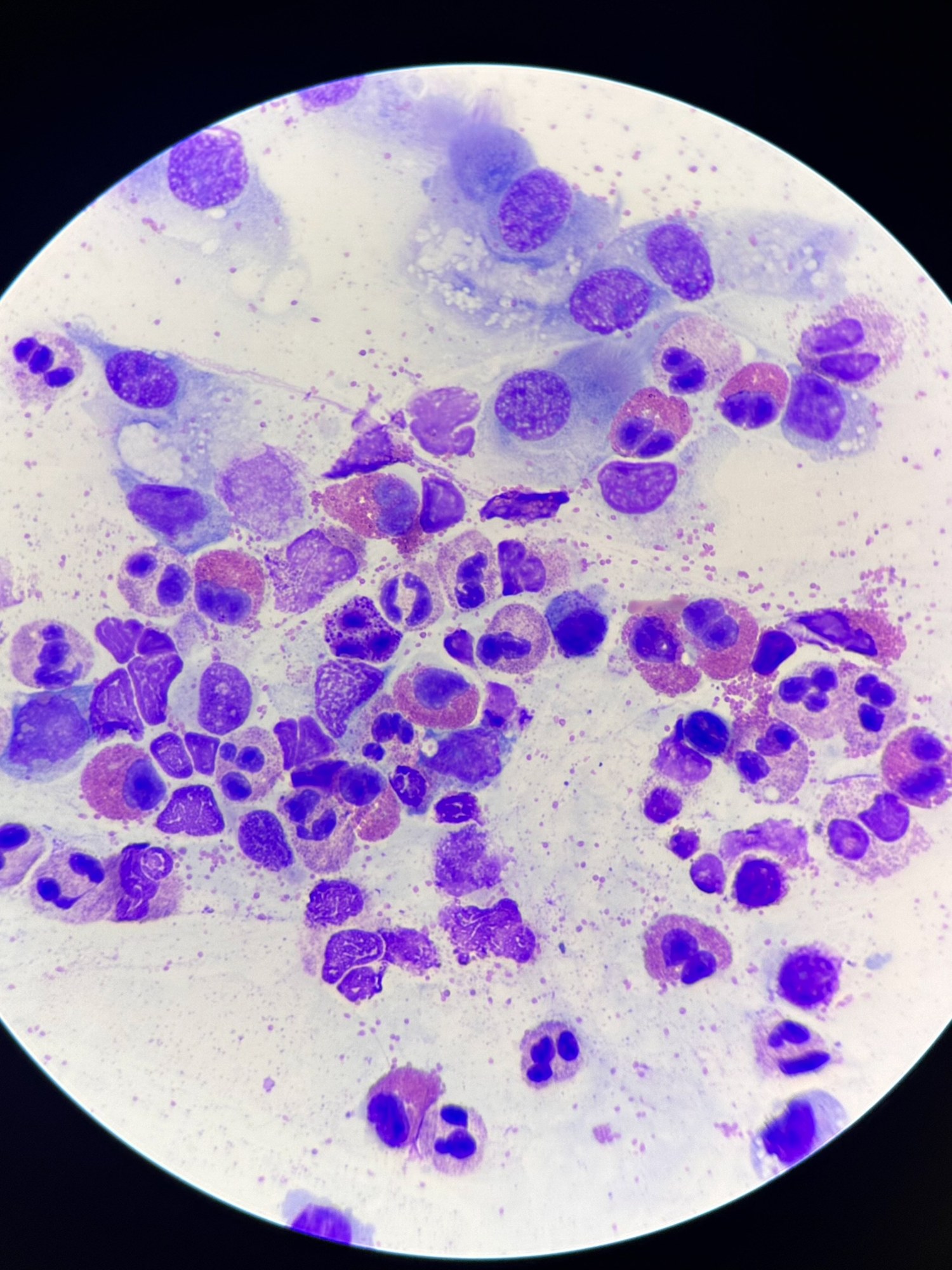
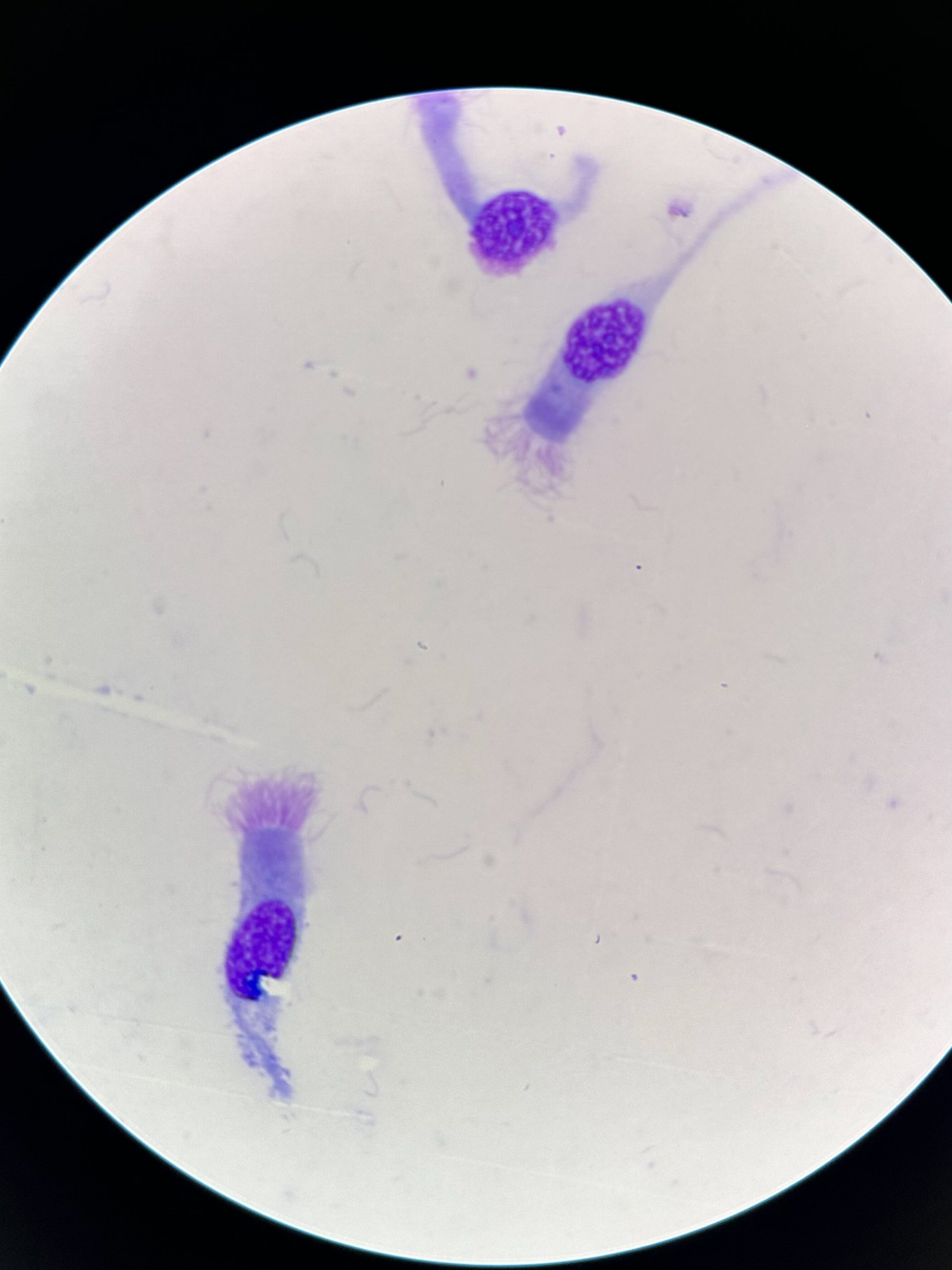
🔬 Cellular composition of nasal secretion. The slide is stained with Romanowsky-Giemsa.
🙏Images kindly provided by clinical laboratory diagnostics physician, Anna Ivanova, Moscow.
📖Reference information: The image shows cells characteristic of nasal secretion, including neutrophils (cells with segmented nuclei), eosinophils (with larger, brightly stained granules), and epithelial cells. The background contains mucin elements and residual protein components of the secretion. The predominance of neutrophils indicates acute inflammation (bacterial infection), while the presence of eosinophils may indicate an allergic reaction.


