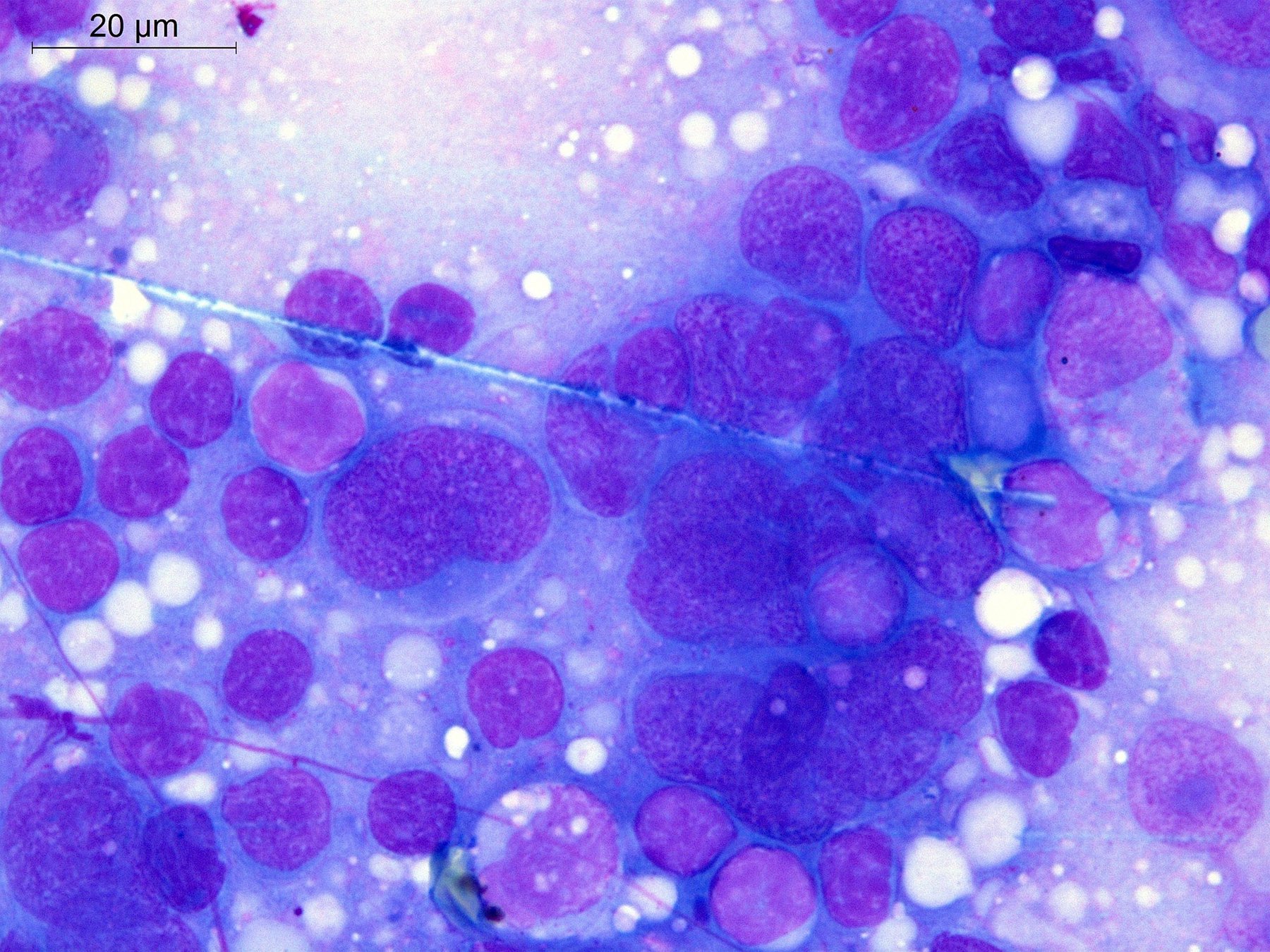
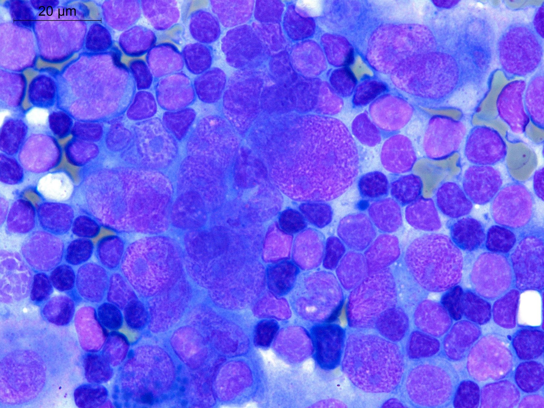
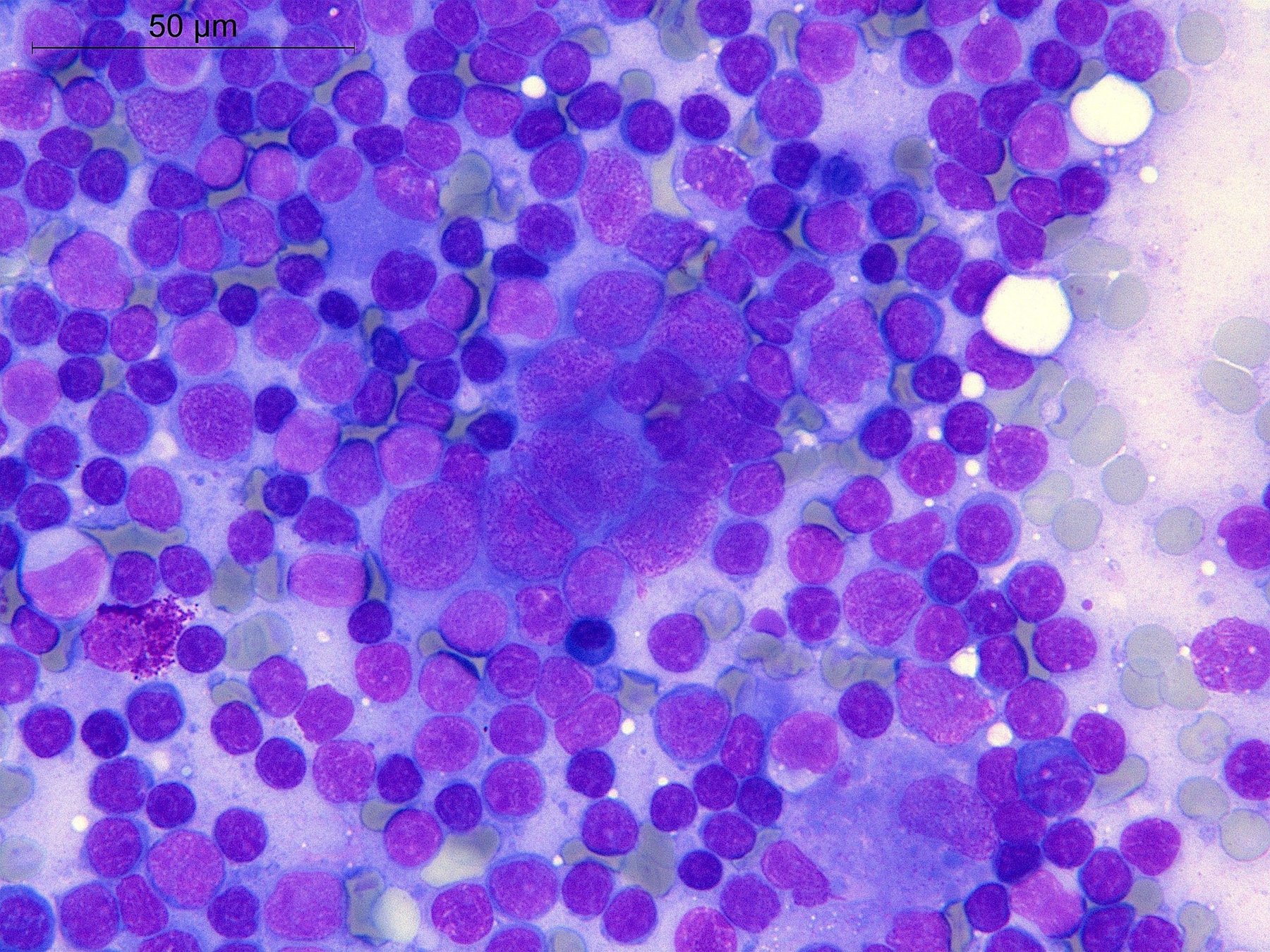
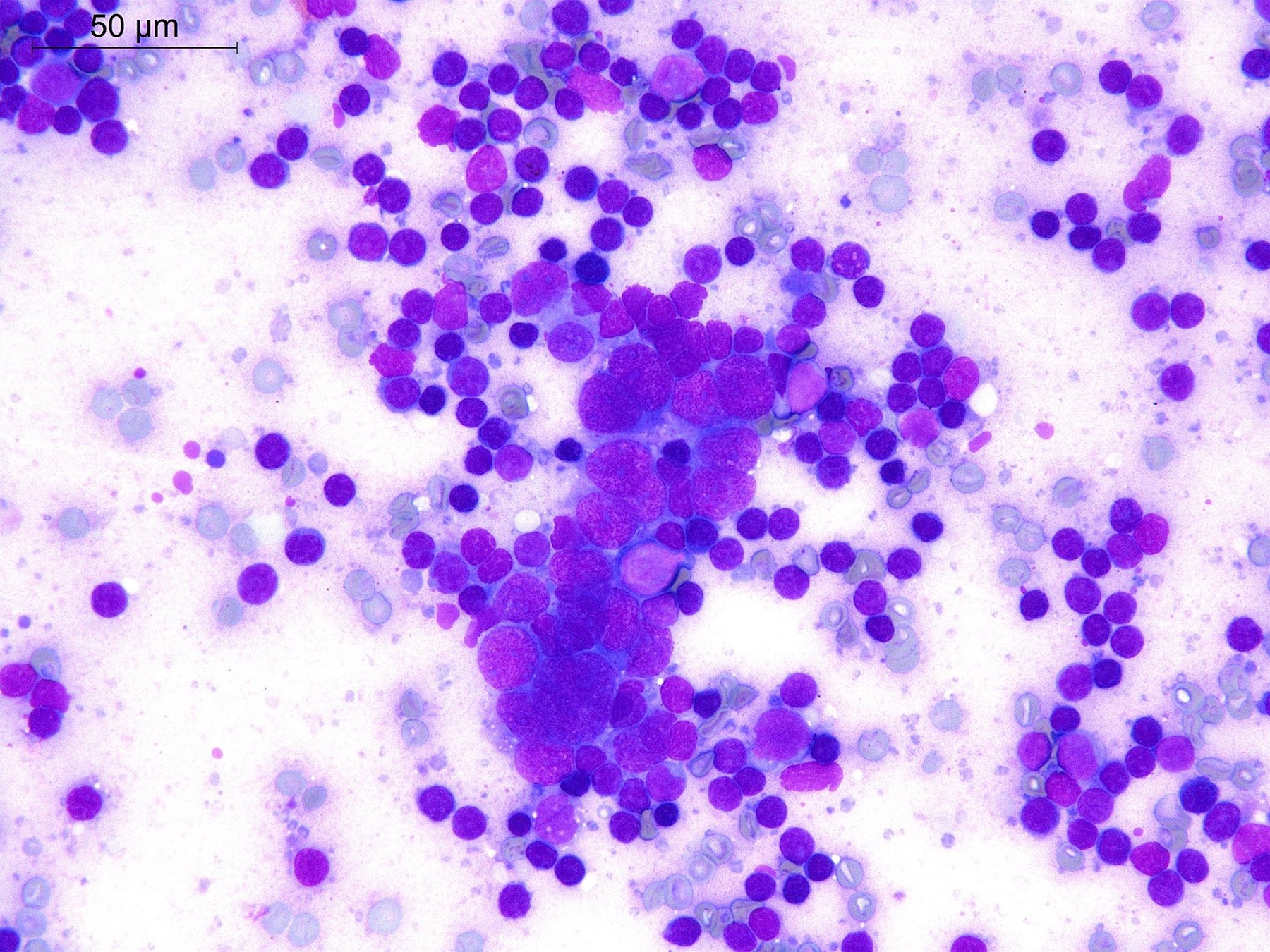
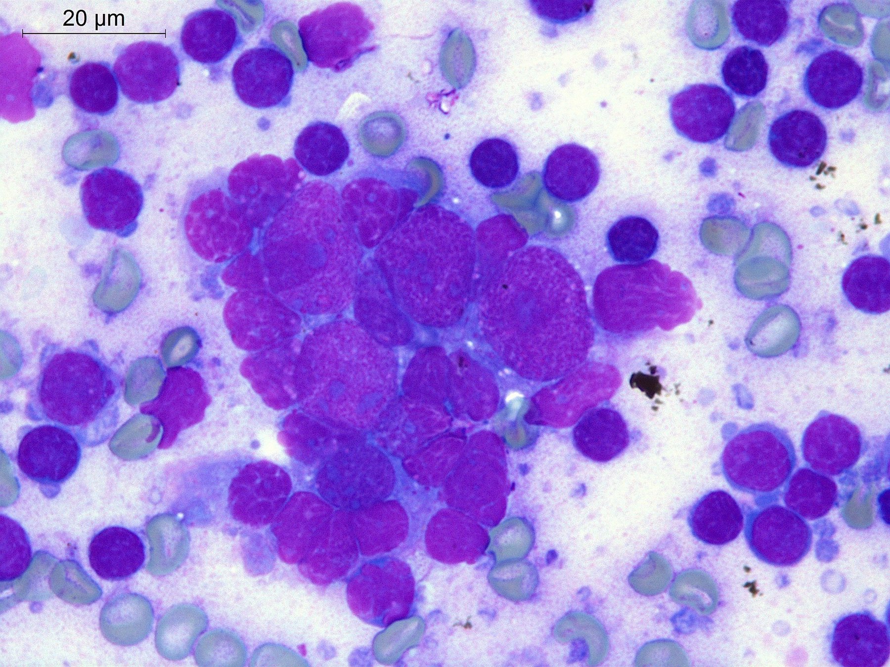
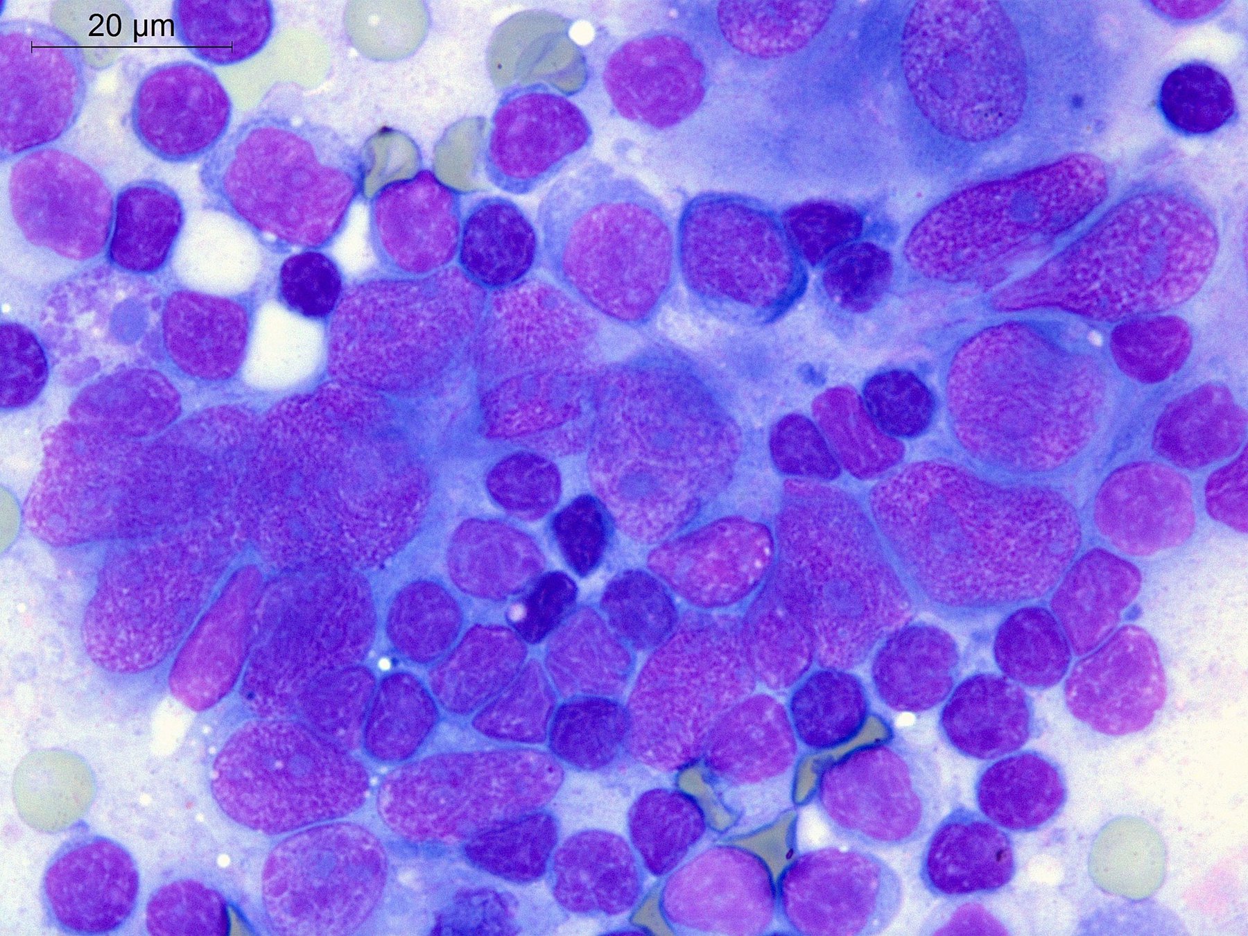
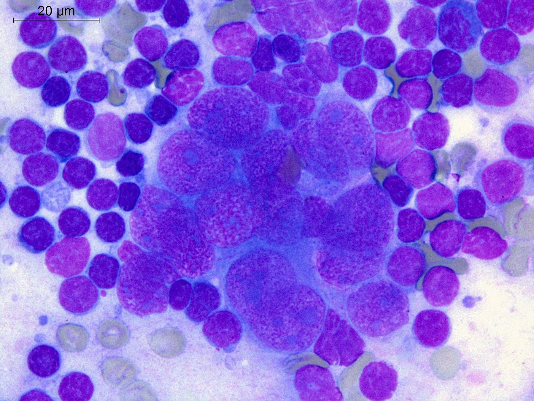
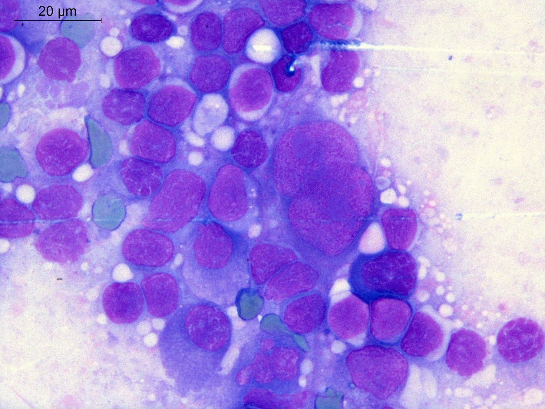
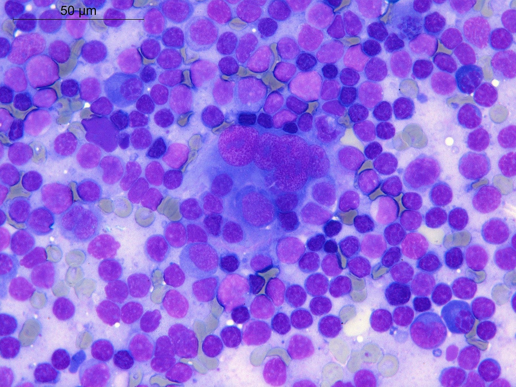
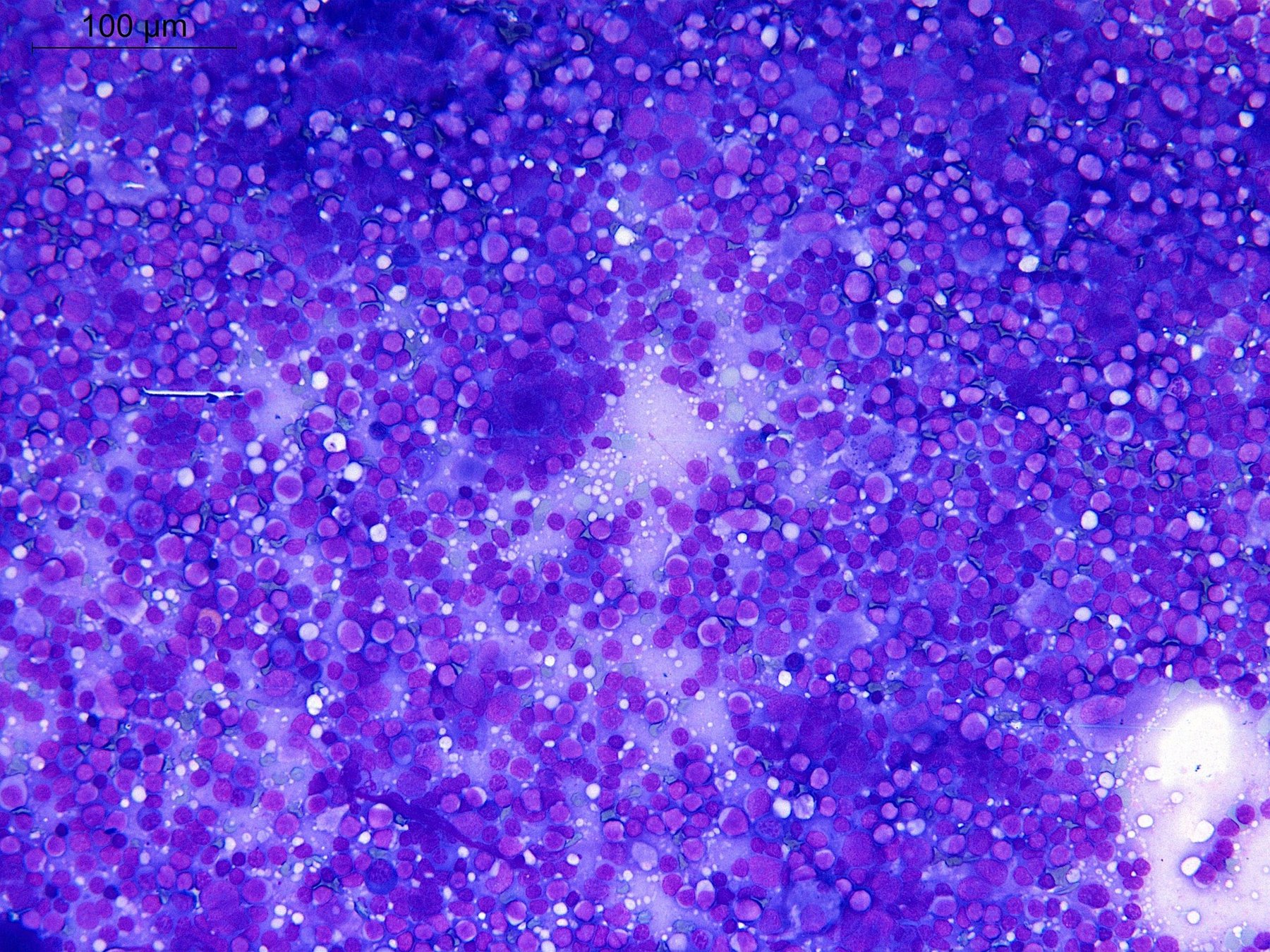
Clinical case of follicular dendritic cell sarcoma. A 63-year-old woman. Excisional biopsy of a cervical lymph node was performed. Based on the results of histological and immunohistochemical studies, follicular dendritic cell sarcoma (FDCS) was diagnosed. Expression of CD21 and CD23 was detected in tumor cells. Ki67 expression level was 30-40%. Expression of pancytokeratin, CK5/6, CD30, CD45, CD68, Sox-10, LMP-1, podoplanin, CD68 was absent in atypical cells. FDCS is a malignant tumor exhibiting morphological and phenotypic characteristics of follicular dendritic cells, which are cells of stromal origin and normally found in germinal centers. FDCS is an extremely rare tumor, difficult for both histological and, even more so, cytological diagnosis. According to the WHO classification, FDCS can mimic a wide range of tumors, such as unclassified pleomorphic sarcoma, meningioma, thymoma, thymic carcinoma, angiosarcoma, undifferentiated carcinoma, lymphoepithelial carcinoma, and gastrointestinal stromal tumor. The diagnostic problem can only be solved by applying a panel of immunohistochemical markers. In the presented material, you can see touch imprints of the lymph node. Cytological diagnosis can be extremely difficult. Typically, these are hypercellular smears in which large tumor cells are visible against a background of small lymphocytes, plasma cells, and histiocytes, arranged both as syncytial sheets and individually. The cytological characteristics of tumor FDCS described in the literature can be variable. Nuclei are round, ovoid, polygonal, and spindle-shaped, and can be tightly apposed or overlap. Nuclear chromatin is vesicular or finely granular, nucleoli are of different sizes. Grooves and pseudoinclusions may be present in the nuclei. Bi- and multinucleated cells are found, sometimes resembling Reed-Sternberg cells. The cytoplasm is medium-sized or wide, pale eosinophilic. In FNA cytological smears, long cytoplasmic processes that form an intersecting network can sometimes be seen. This is not visible in our preparation, as it is not an aspirate smear, but a tissue imprint. Mitoses, pronounced nuclear atypia, and necrosis may be present. 👉 The clinical case was kindly provided by the laboratory physician, pathologist, member of the FLM hematology committee Elena Belyakova, Department of Molecular Morphology, E.E. Eichwald Clinic, I.I. Mechnikov Northwestern State Medical University, St. Petersburg.
Follicular Dendritic Cell Sarcoma: Cytology of a Rare Tumor
Cytological features and immunophenotype of follicular dendritic cell sarcoma (FDCS) of a cervical lymph node: a clinical case description.