

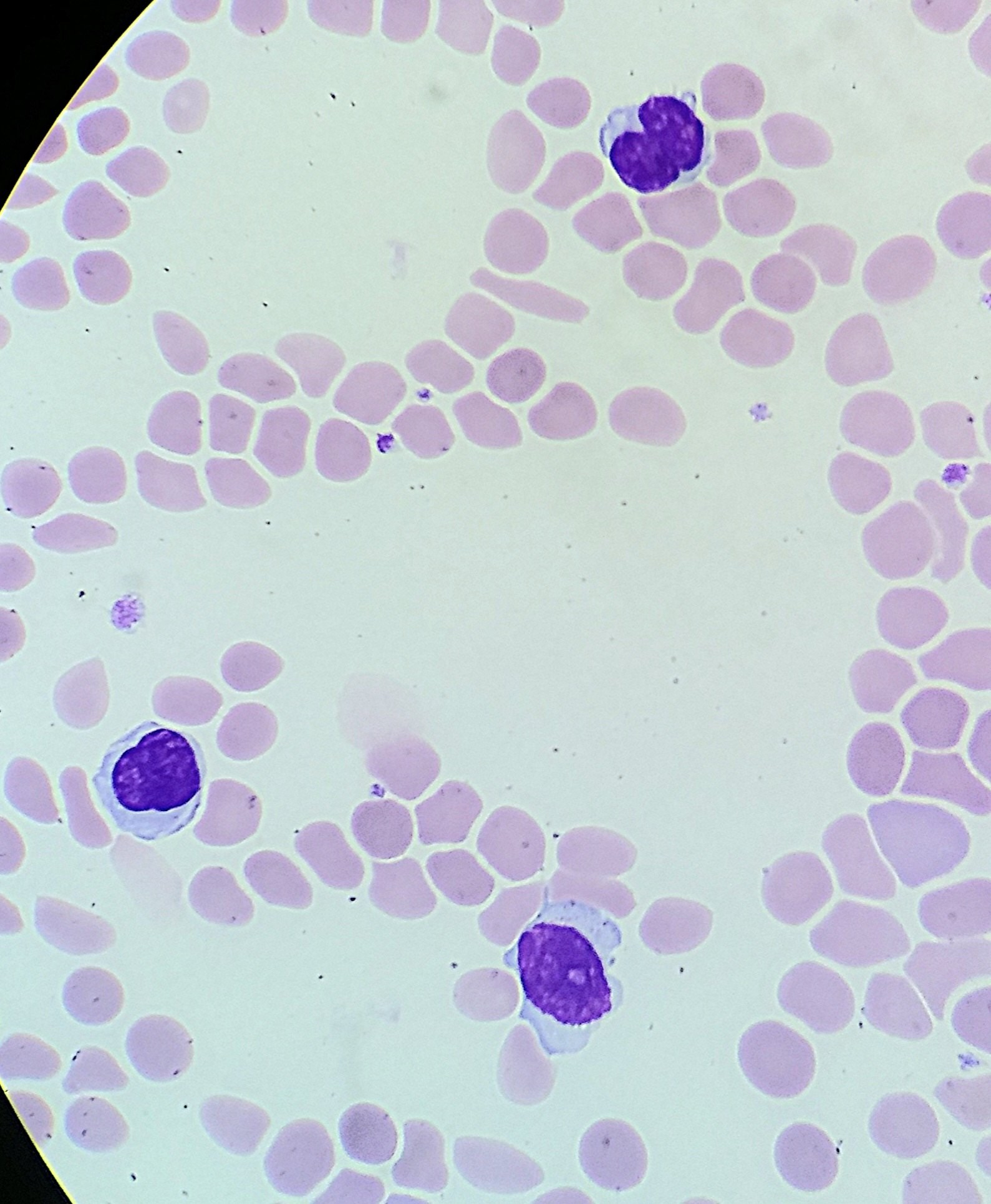
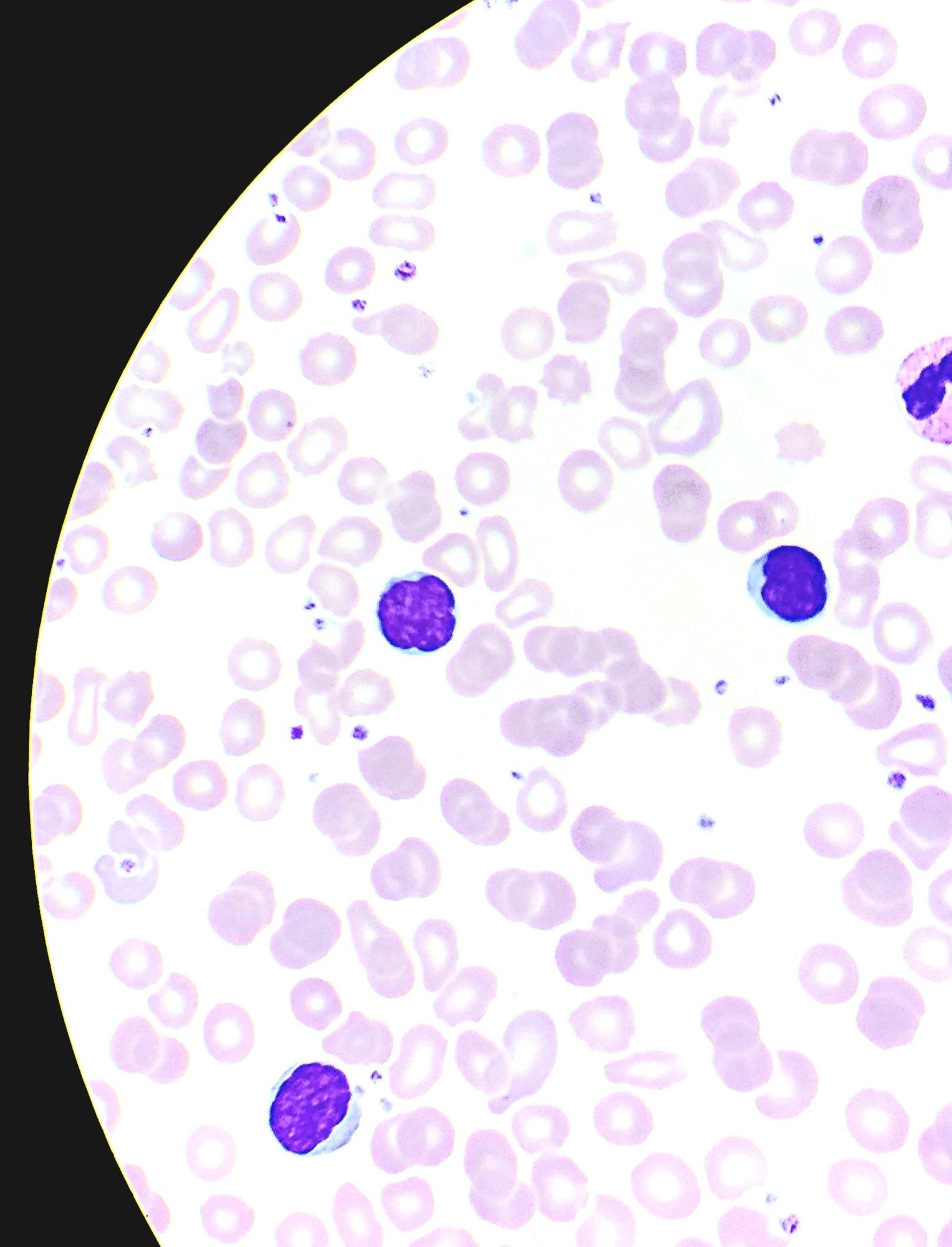
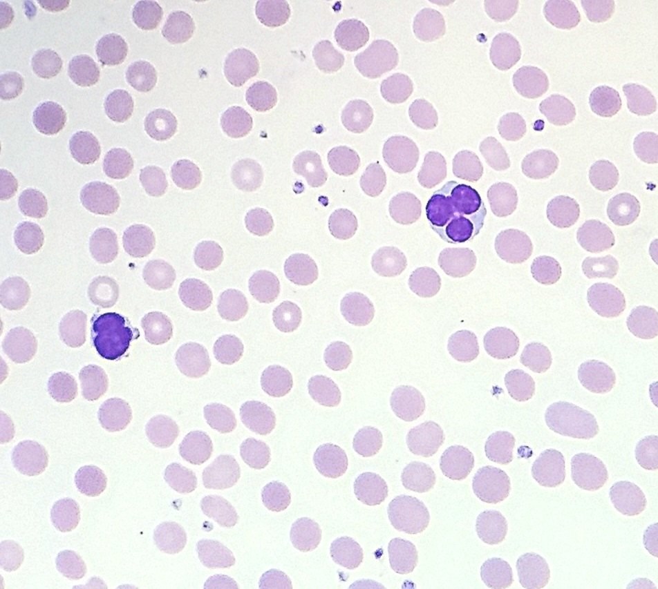
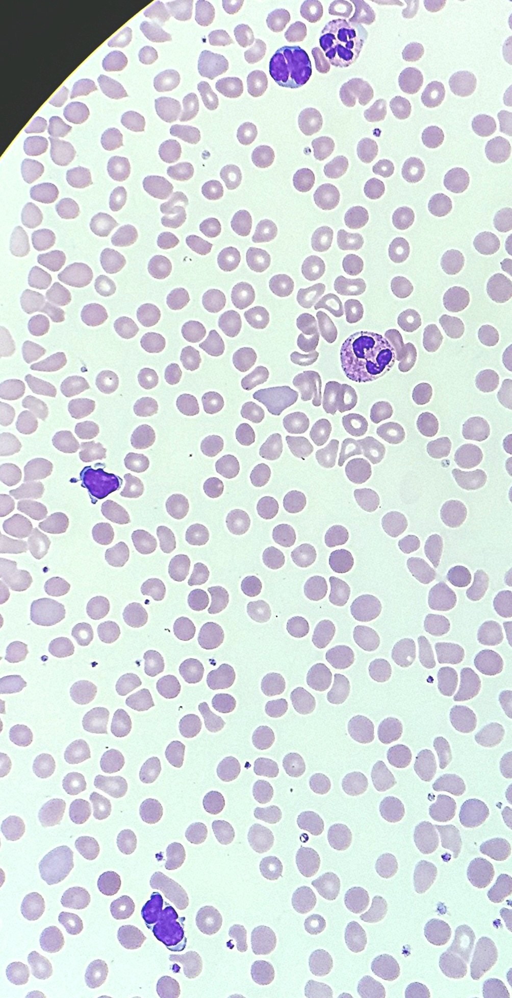
Sézary Syndrome. A clinical case from 🇮🇱 Israel. ✅ A 79-year-old patient complains of skin rashes, general weakness, and weight loss for about 1.5 months.
🩸 In peripheral blood, absolute lymphocytosis is 8.5 thousand/µL, hypochromic anemia is 87 g/L, and platelet (PLT) count is within normal limits.
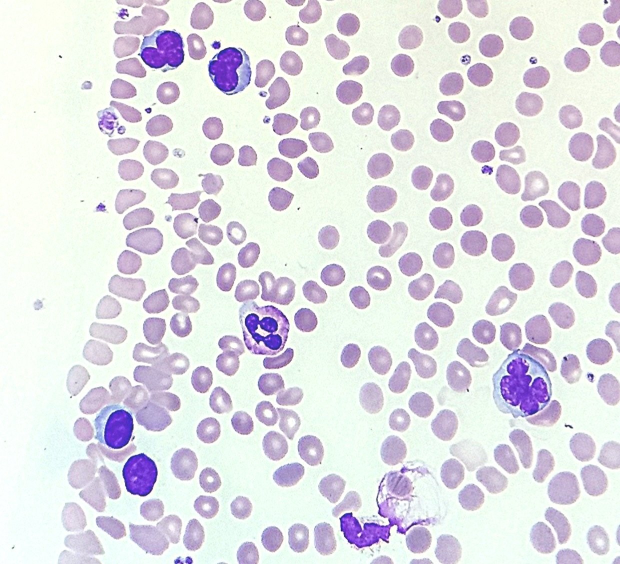
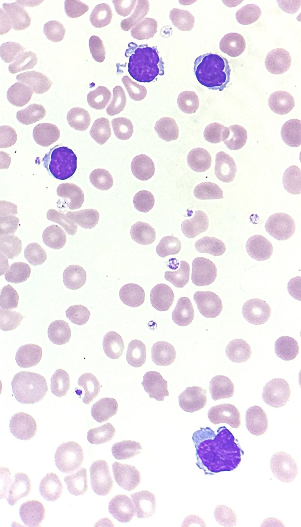
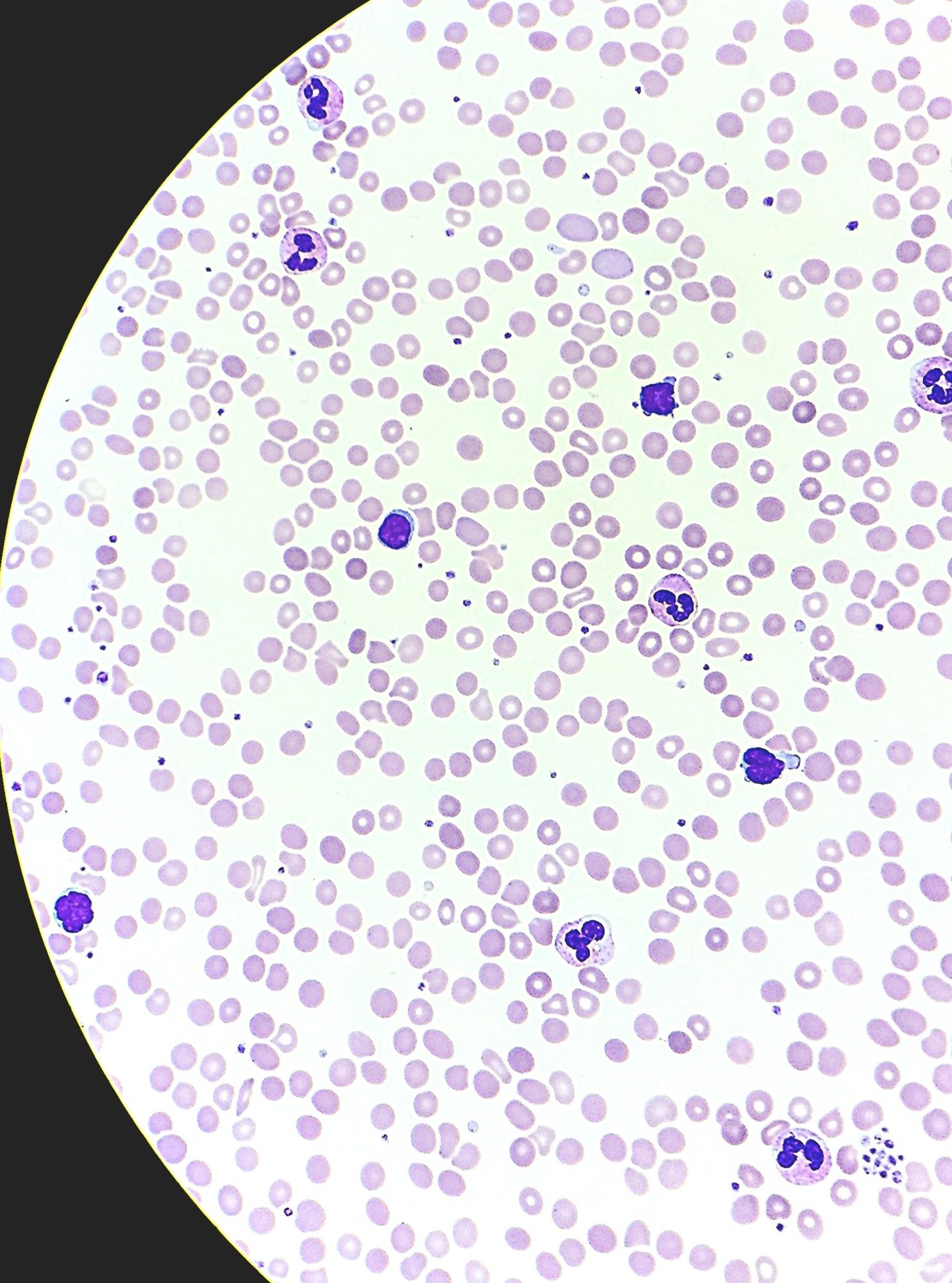
🔬 In the peripheral blood smear, lymphoid cells with hyperchromatic nuclei are observed, appearing cleaved and convoluted. The characteristic “cerebriform” structure is not particularly pronounced. However, it is important to remember that this can also occur.
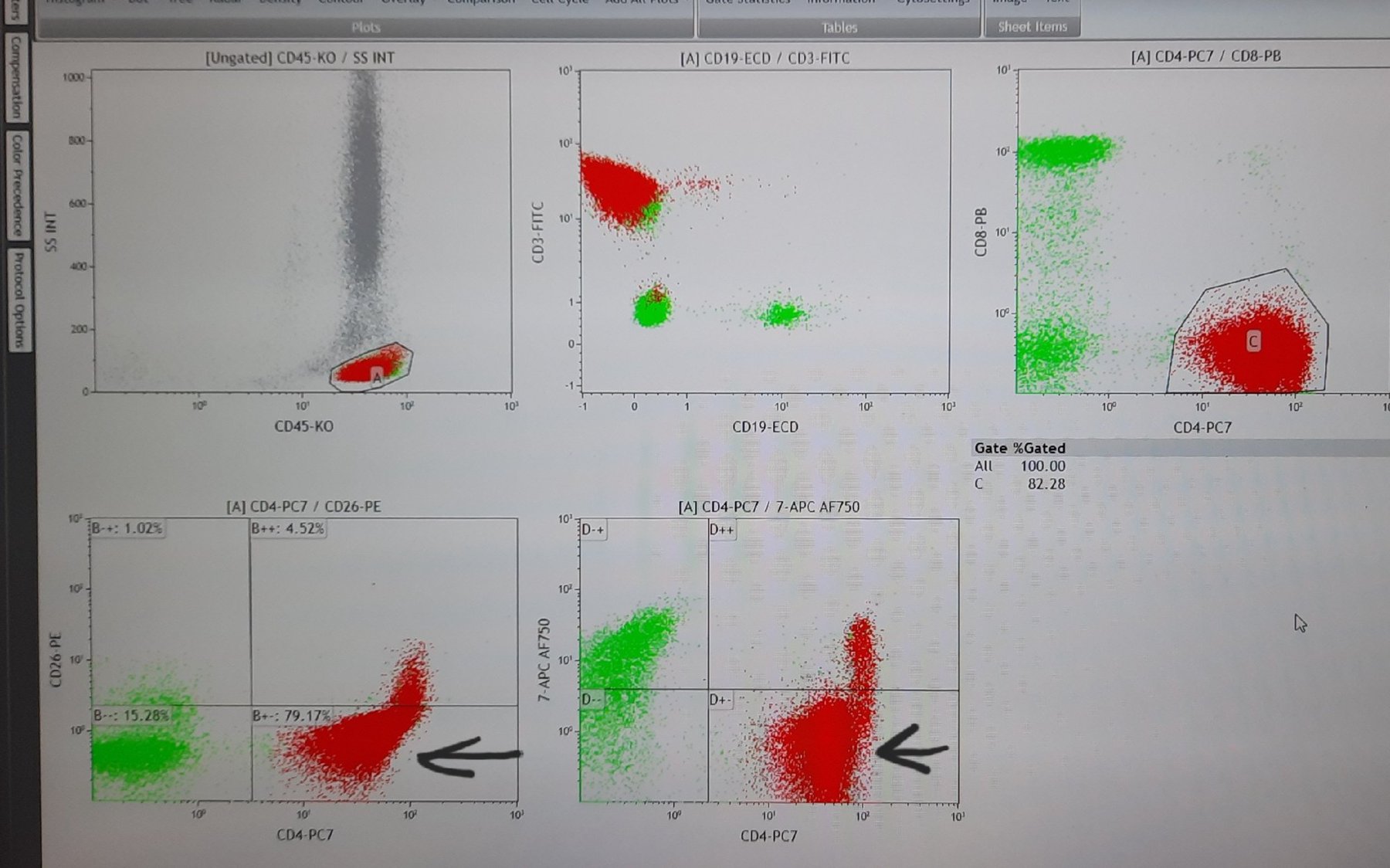
💡 Typical picture on flow cytometry: CD4=82%, the CD4+/CD7-/CD26- population constitutes almost 80%. ✅ Molecular genetic analysis result: TCRG-rearrangement: positive (monoclonal).
🔬Case from the Head of the Morphology/Flow Cytometry Department of HaEmek Medical Center, Afula 🇮🇱Israel - Anna Belen.


