

Temporal Arteritis or COVID-19? The Usefulness of the ESR Test. Case #1: A 70-year-old female presented to the emergency department with complaints of throbbing headache and blurred vision. She stated the headache started 2 days ago, initially in her temples, but had worsened over the last few hours. Complete blood count results were unremarkable except for an elevated platelet count of 480 K/µL (normal 150-450 K/µL). ESR 110 mm/hr (normal up to 15 mm/hr) CRP was 2.51 mg/L (normal up to 5 mg/L).
The patient was scheduled for a temporal artery biopsy with suspicion of temporal arteritis, also known as giant cell arteritis (GCA). Temporal arteritis is an autoimmune disease that causes inflammation of the temporal arteries. Under the microscope, the inflamed cells of these arteries appear giant, hence the name of the disease. The inflammation causes narrowing of the arteries, can affect chewing and eating, and can lead to blindness if left untreated. The treatment of choice is corticosteroids, often prescribed for at least a year. Symptoms are often controlled, and laboratory results, including ESR, can be used to monitor the condition and response to treatment.
If you are still wondering if ESR should be discontinued as a useful test, patients with COVID infection and elevated ESR are now being observed. Over the past 2 years, several articles have been written about elevated ESR in COVID-19 patients. One study aimed to evaluate the usefulness of ESR in distinguishing between severe and non-severe cases of COVID-19. The study suggests that severe cases of COVID-19 are associated with a higher elevation of ESR compared to non-severe cases. High ESR persisted for a long time even after the patient recovered from COVID-19, with no other inflammatory processes or other conditions causing ESR elevation found.
Case #2: My second case is that of a 58-year-old female with ear pain and a throbbing temporal headache. An ear infection was ruled out, and the patient was referred to an ophthalmologist to rule out temporal arteritis. The patient’s CRP was elevated, but her ESR and platelet count were within normal limits. The patient underwent a COVID test as part of a pre-operative workup before a temporal artery biopsy, and the COVID-19 test was positive. Over the past year, there have been cases in the literature of this new set of symptoms in COVID-19 patients. The conclusion from these cases is that if a patient presents with symptoms consistent with temporal arteritis, with an elevated CRP, but with normal ESR and platelet levels, then patients should be tested for COVID-19.
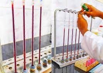
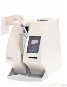
ESR is one of the oldest laboratory tests still in use. The study of blood sedimentation was one of the principles upon which ancient Greek medicine was based. In the 1700s, doctors noticed that the rate of erythrocyte sedimentation changed during illness. This theory was first introduced as a laboratory test over 100 years ago. Depending on the historical accounts and articles you read, it was first described by the Polish physician Edmund Biernacki in 1897 or the Swedish physician Robert Fåhraeus in 1915. Biernacki proved the connection between sedimentation rate and fibrinogen levels in the blood and proposed using ESR in diagnosis. Alf Vilhelm Albertsson Westergren (a familiar name!) in the early 1920s went even further, developing a blood collection technique and defining standards for ESR. To this day, the Westergren method for determining erythrocyte sedimentation rate is recognized as the gold standard reference method for ESR measurement. Erythrocyte sedimentation rate measures the speed at which red blood cells settle under gravity, in mm/hour. Red blood cells usually repel each other due to zeta potential, and aggregation is inhibited. In conditions with increased fibrinogen or immunoglobulins, these proteins coat red blood cells, promoting their aggregation. In conditions such as anemia, ESR will be high because with a lower hematocrit, the rate of upward plasma flow changes, and erythrocyte aggregates fall faster. In polycythemia, increased blood viscosity can cause a decrease in ESR. In sickle cell anemia and other conditions such as spherocytosis, red blood cells have an abnormal shape, which reduces ESR.
ESR is a simple, inexpensive, non-specific test that has been used for many years to diagnose conditions associated with acute and chronic inflammation. An elevated ESR is not associated with a specific diagnosis; therefore, it must be used in conjunction with other tests. Conversely, a normal ESR cannot be used to rule out the presence of a serious disease. ESR should also not be used as a screening test in asymptomatic patients. Since fibrinogen is an acute phase protein, ESR is elevated in many inflammatory and neoplastic conditions accompanied by increased fibrinogen levels, including diabetes, infections, pelvic inflammatory disease, lupus, rheumatoid arthritis, acute coronary syndrome, and neoplasms. However, non-inflammatory factors such as old age, female sex, and pregnancy can also cause an elevated ESR.
Historically, ESR has been used to identify inflammatory conditions and monitor disease progression or response to treatment. More specific tests have been developed for many of these conditions, but ESR still has its advantages. Interestingly, for a test that was on the “obsolete” list 12 years ago, over 50 articles in scientific journals have been written about ESR in the last 2 years. ESR can rule out unnecessary tests and help reduce medical costs. It has its advantages in small laboratories and rural areas, as it can provide quick results without expensive equipment. For laboratories that do not perform more complex tests such as CRP and procalcitonin, ESR can provide answers without waiting for results from central laboratories. Although ESR can take 1 hour, it is much faster than sending biomaterial for testing to a central laboratory. Source: labmedicineblog.com/category/hematology/


