

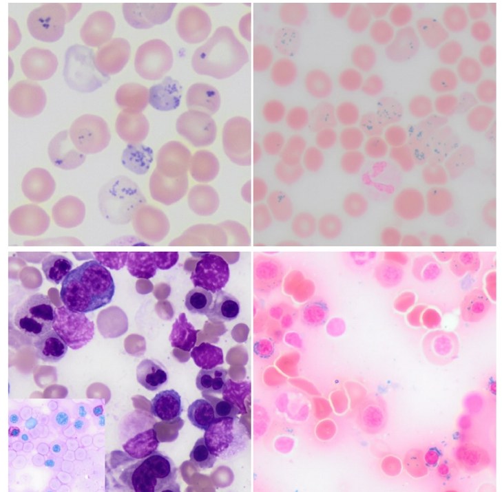
🔬 Atypical Pappenheimer bodies in myelodysplastic syndrome: how not to confuse them with basophilic stippling?
Unusual Pappenheimer bodies 🩸 were found in the blood smear of a patient with myelodysplastic syndrome (MDS) and pulmonary embolism (PE).
📌 What is the difficulty? These inclusions in erythrocytes externally resembled basophilic stippling (a classic sign in lead poisoning or thalassemia). However, a detailed analysis revealed:
🔹 Iron-containing granules (Pappenheimer bodies) — a manifestation of impaired iron utilization in MDS. 🔹 An erroneous diagnosis could lead to overdiagnosis of other conditions.
💡 Why is this important? ✅ Differential diagnosis is critical: MDS requires a special approach to treatment. ✅ PE in MDS is a common complication due to hypercoagulation.
Conclusion: Smear microscopy is a powerful tool, but requires attention to detail! 🔍
Source: Chen S., Zheng Q. Atypical Pappenheimer bodies mimicking basophilic stippling in myelodysplastic syndrome with pulmonary embolism. Br J Haematol. 2025;207:13-14.


