

🔬 Howell-Jolly bodies in every field of view, +++. Female, 68 years old.
🙏 Photos kindly provided by Clinical Laboratory Diagnostics Physician, Irina Selivanova LLC “Citilab-Ural”, Yekaterinburg
A clinical case of Howell-Jolly bodies in erythrocytes (+++) in a 68-year-old female patient. Morphology of inclusions in blood smear and hematology analyzer data.
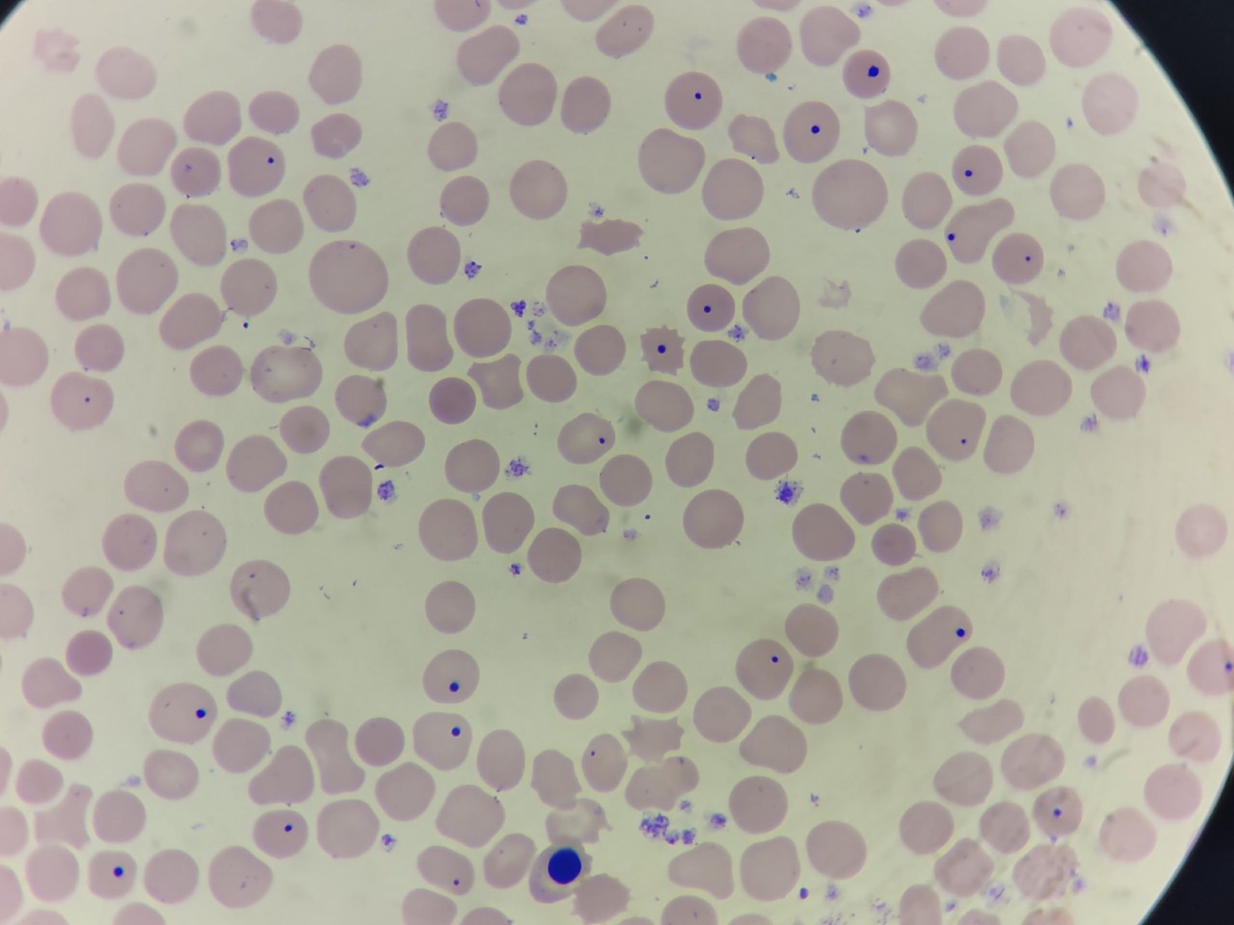
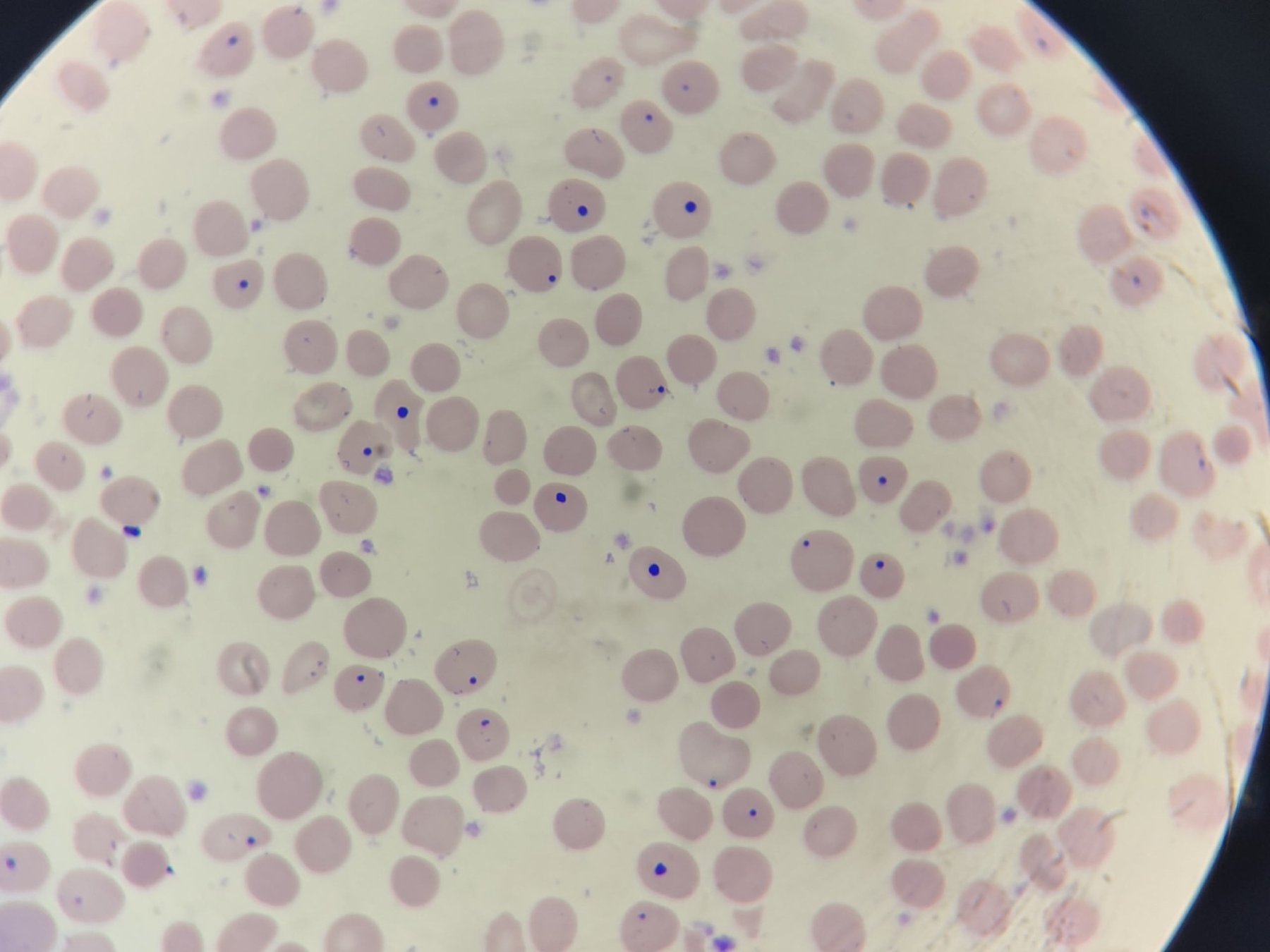
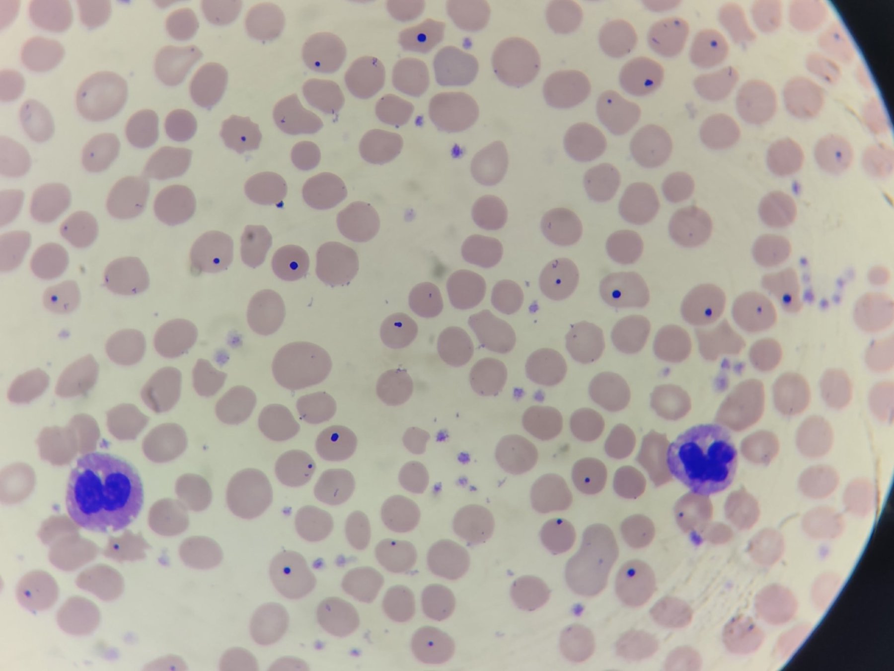
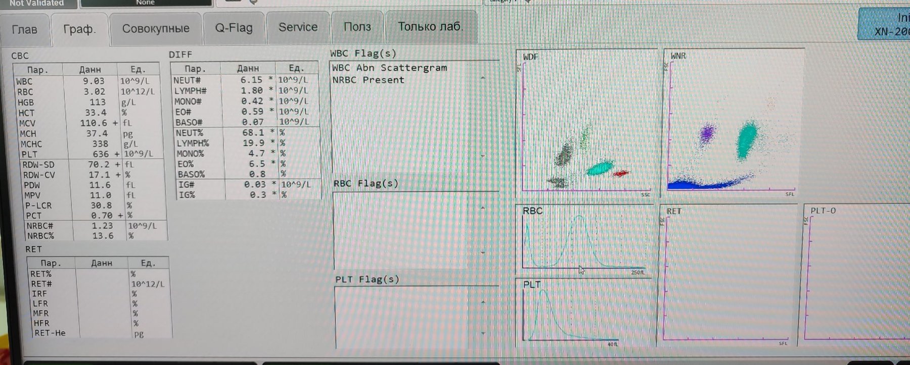
🔬 Howell-Jolly bodies in every field of view, +++. Female, 68 years old.
🙏 Photos kindly provided by Clinical Laboratory Diagnostics Physician, Irina Selivanova LLC “Citilab-Ural”, Yekaterinburg

A case of secondary erythremia in a male patient: microcytosis, Howell-Jolly bodies in erythrocytes, and evaluation of hematology analyzer histograms and numerical parameters.

Erythrocyte morphology in newborns: detection of Howell-Jolly bodies and binucleated normoblasts in peripheral blood, causes, and evaluation criteria.

Morphology of reticulocytes, methods of supravital blood smear staining, reference ranges, causes of reticulocytosis and reticulocytopenia in clinical practice.