

🔬 Howell-Jolly bodies in a newborn infant. Peripheral blood. Romanowsky-Giemsa stain, x100 objective
🙏 Photos and description kindly provided by biologist Anna Feditskaya, Luhansk
Morphology of erythrocytes with Howell-Jolly bodies in a neonatal peripheral blood smear. Microscopy with immersion oil after Romanowsky-Giemsa staining.
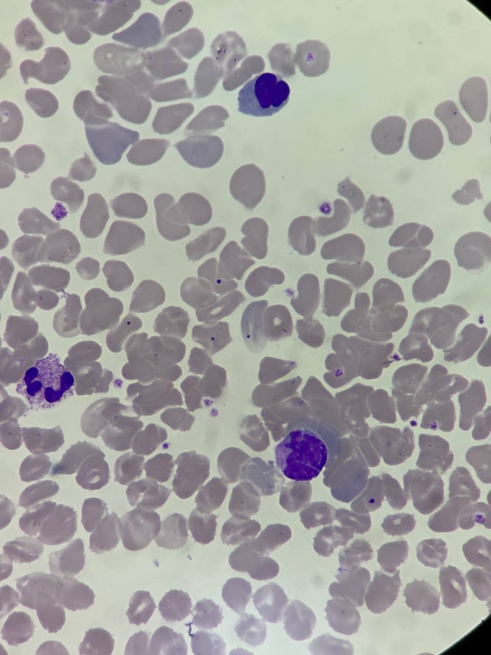
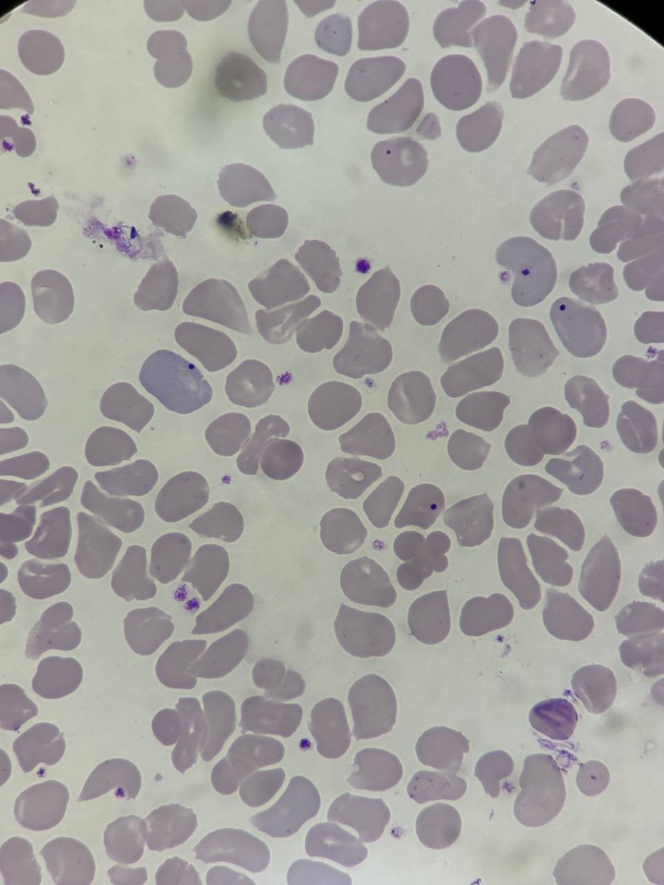
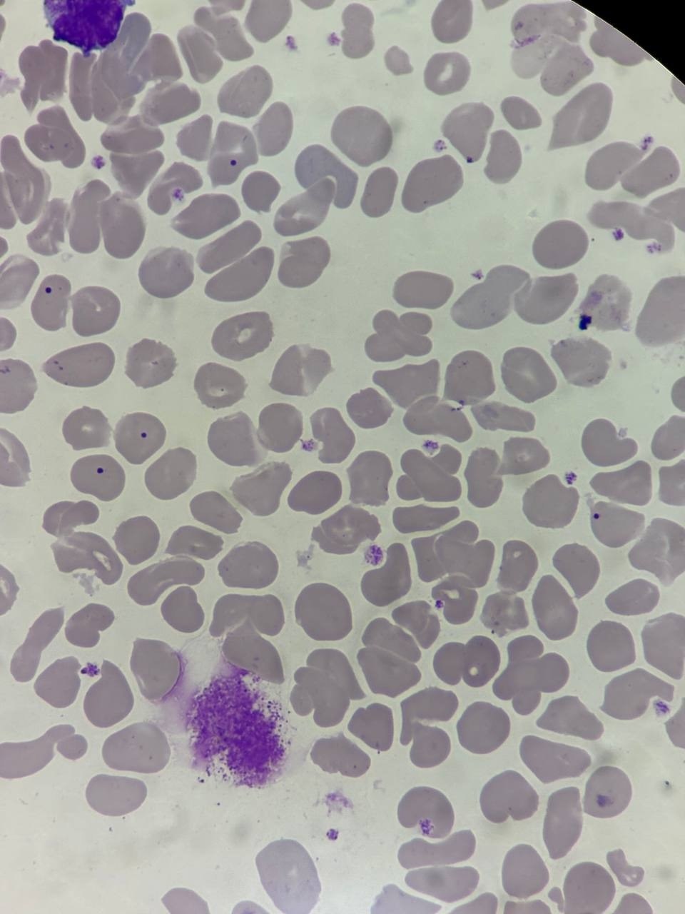
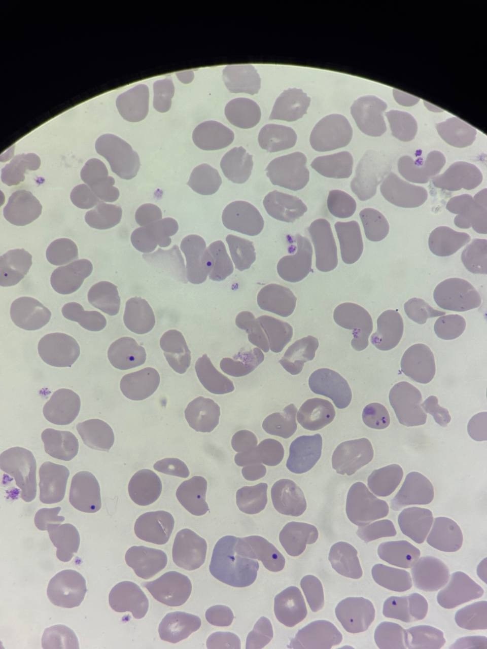
🔬 Howell-Jolly bodies in a newborn infant. Peripheral blood. Romanowsky-Giemsa stain, x100 objective
🙏 Photos and description kindly provided by biologist Anna Feditskaya, Luhansk

What are Howell-Jolly bodies in a blood smear? Learn about the morphology of nuclear inclusions in red blood cells and their causes in asplenia, splenectomy, and hemolysis.

What are Howell-Jolly bodies in erythrocytes? Learn about the mechanism of nuclear remnant formation and their causes in megaloblastic anemia, hemolysis, and asplenia in blood smears.

Morphology of pathological inclusions in erythrocytes: Cabot rings and Howell-Jolly bodies in peripheral blood smear in erythropoiesis disorders.