

🔬 Microscopy of a female smear for flora: Trichomonas Vaginalis.
🙏 The publication was kindly provided by medical laboratory technician Nafisa Khisamutdinova, Surgut
Learn about Trichomonas vaginalis morphology in gynecological smears. This guide covers differential diagnosis of trichomonads, epithelial cells, and co-occurring microflora.
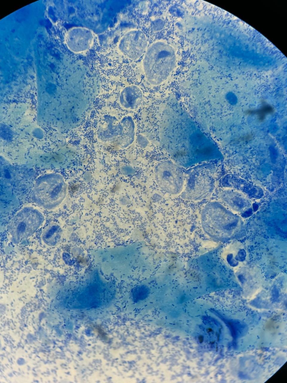
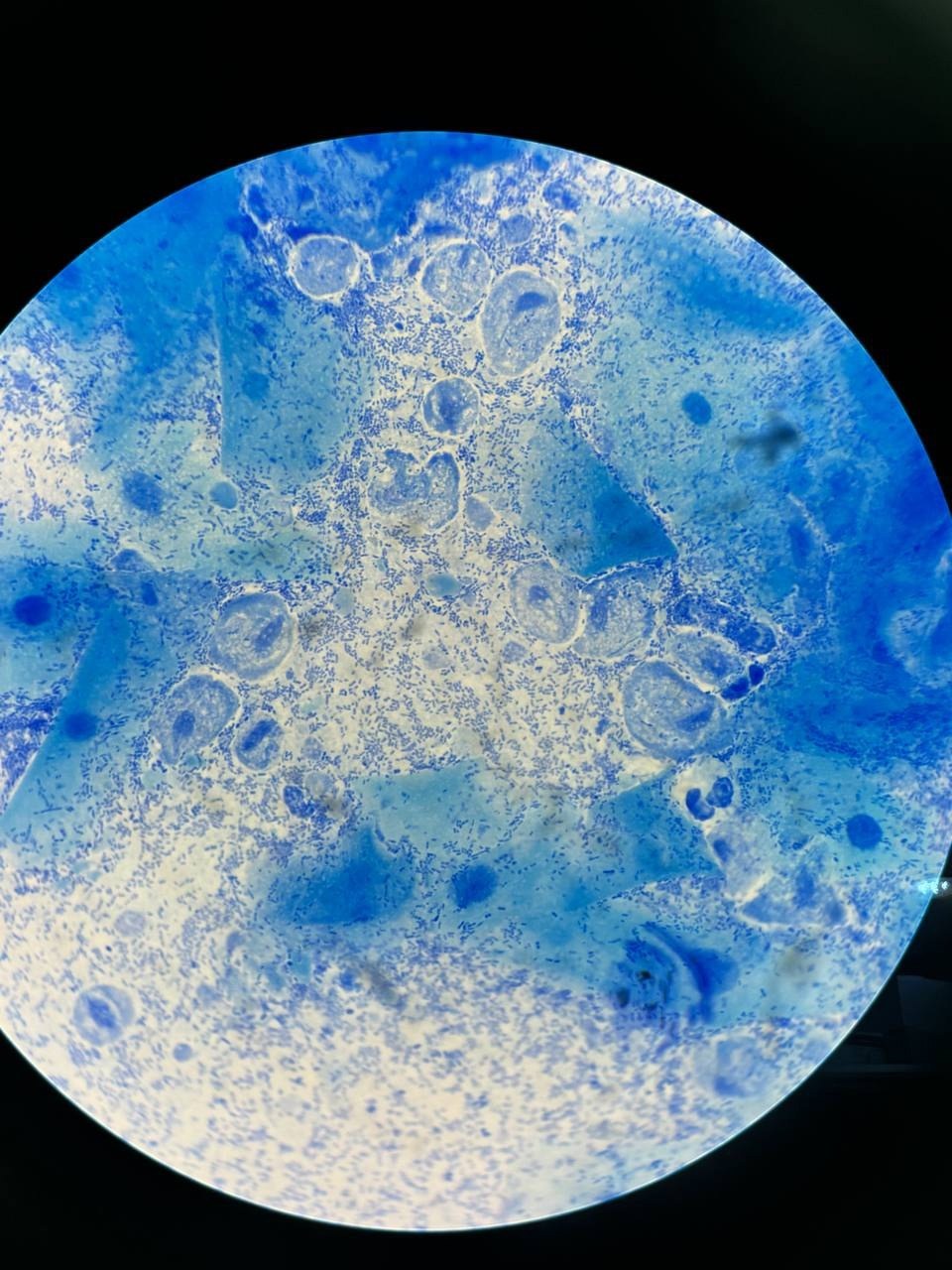
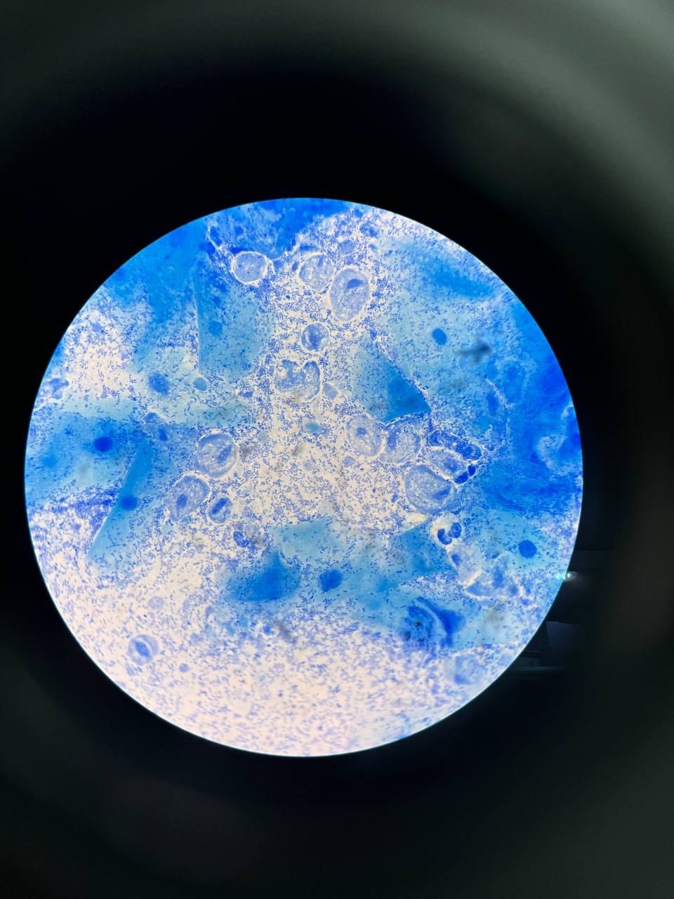
🔬 Microscopy of a female smear for flora: Trichomonas Vaginalis.
🙏 The publication was kindly provided by medical laboratory technician Nafisa Khisamutdinova, Surgut

A clinical case of detecting Trichomonas vaginalis and massive leukocytosis in a genital tract smear from a pregnant patient at 38 weeks.

Microscopic appearance of Trichomonas vaginalis in a stained male flora smear: leukocyte reaction, epithelial cells, and PCR confirmation.

Morphology of Trichomonas vaginalis in stained urogenital smears: identification of protozoa, leukocyte reaction, and bacterial flora during microscopy.