

🔬 Trichomonas vaginalis in vaginal discharge smear. 1,2,3 photos - methylene blue stain, x1000 4,5 photos - Romanowsky stain, x1000
🙏 Material for publication kindly provided by: Elizaveta Emelyanova, Rostov-on-Don, Laboratory Diagnostics Physician
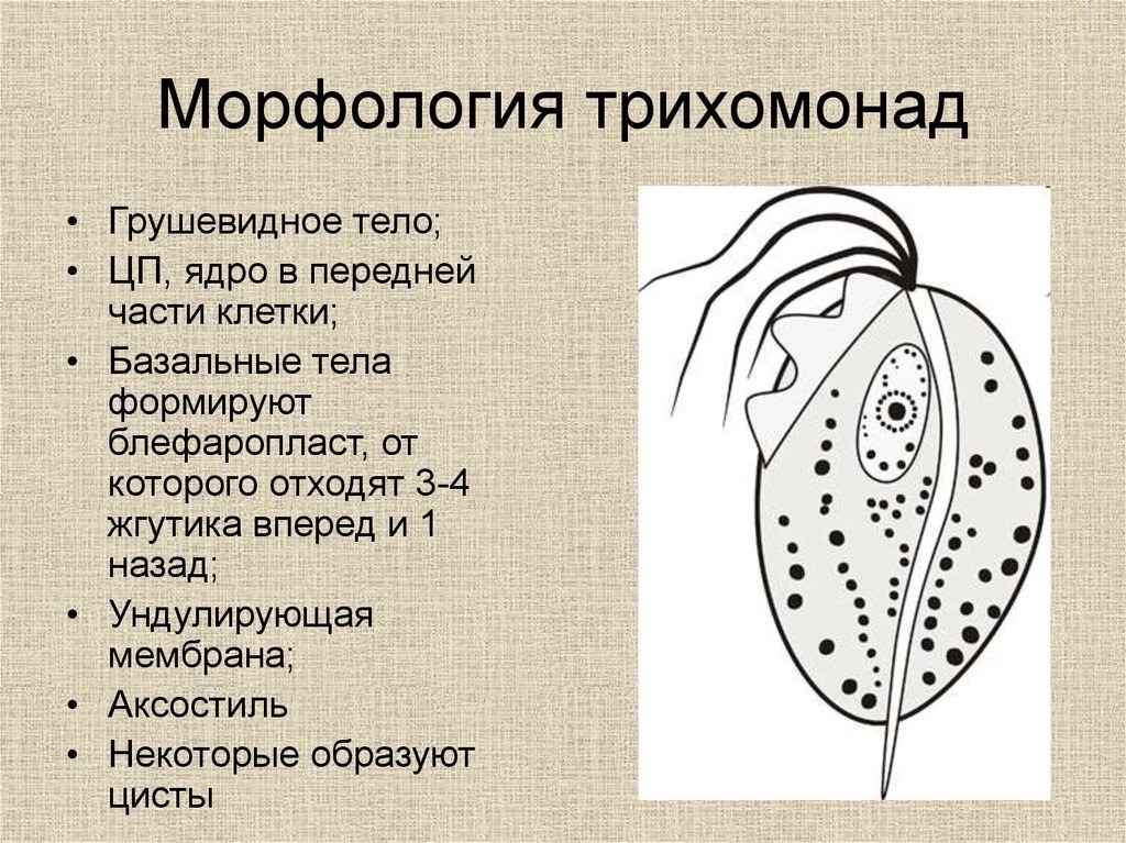
Microscopic diagnosis of Trichomonas vaginalis in vaginal discharge smears: parasite morphology with methylene blue and Romanowsky staining.
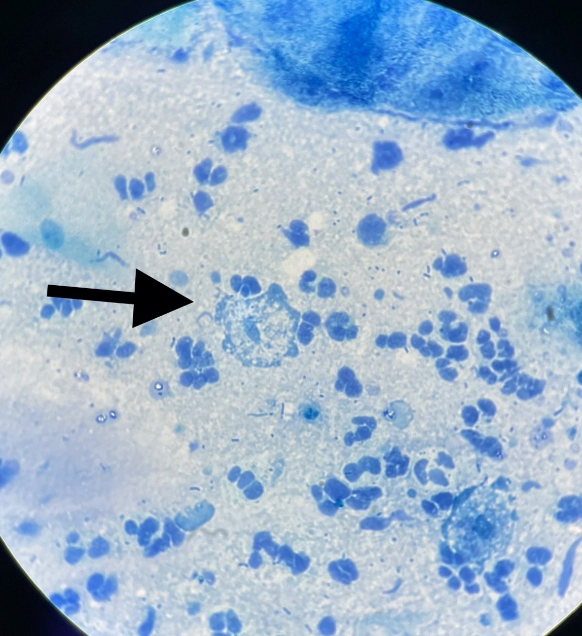
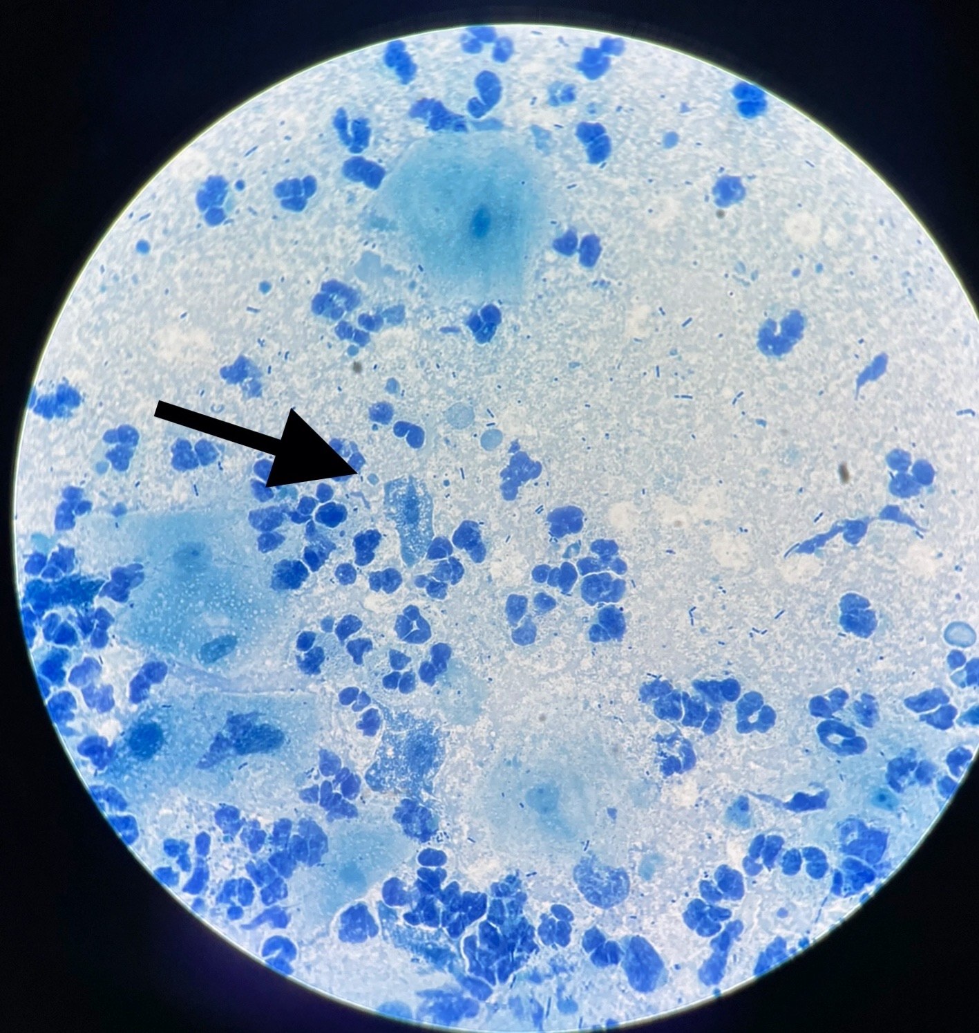
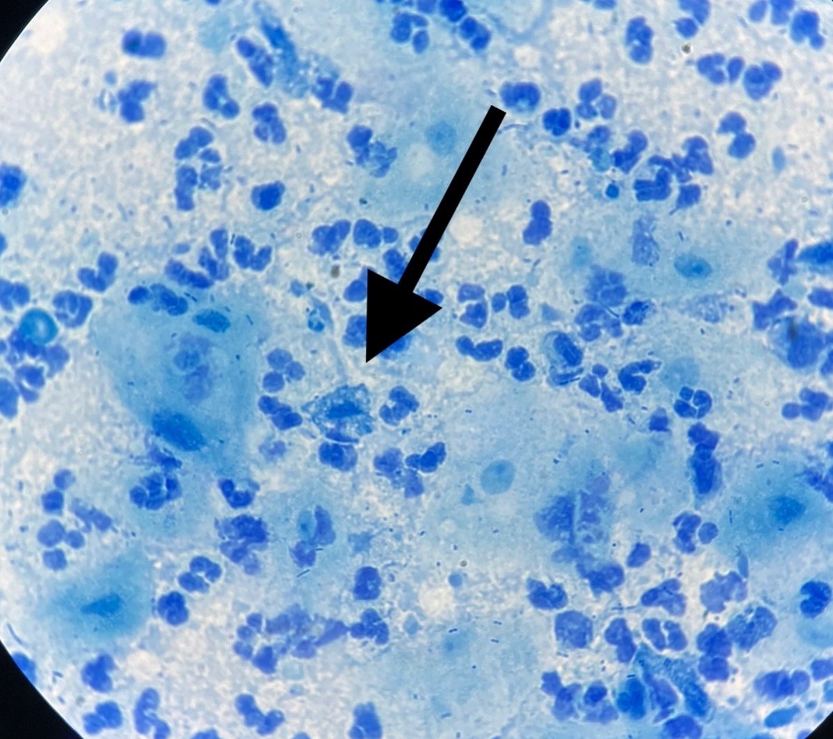
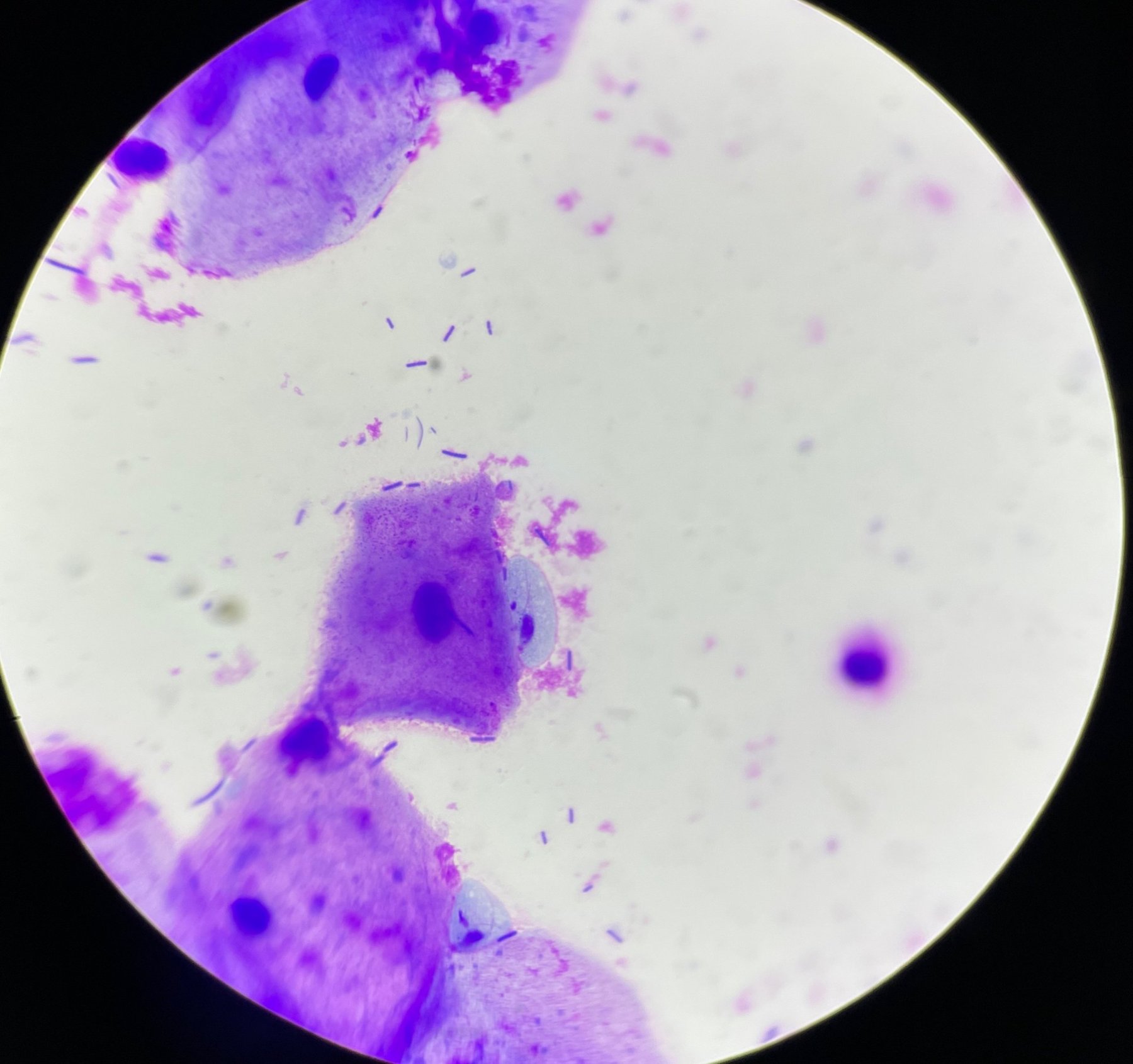
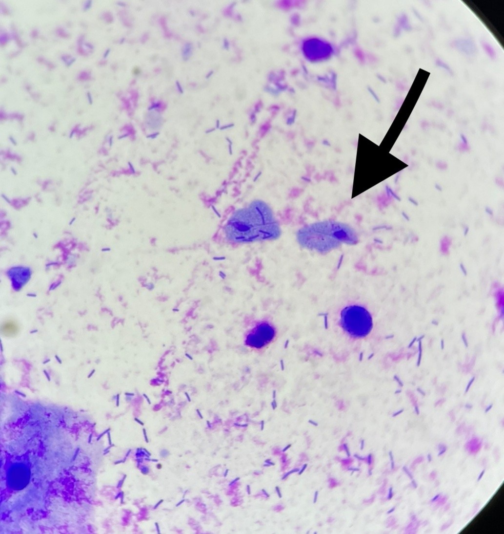
🔬 Trichomonas vaginalis in vaginal discharge smear. 1,2,3 photos - methylene blue stain, x1000 4,5 photos - Romanowsky stain, x1000
🙏 Material for publication kindly provided by: Elizaveta Emelyanova, Rostov-on-Don, Laboratory Diagnostics Physician

Microscopic view of a urogenital smear: identifying Trichomonas vaginalis trophozoites among squamous epithelial cells and abundant microflora.

Microscopic appearance of Trichomonas vaginalis in a stained male flora smear: leukocyte reaction, epithelial cells, and PCR confirmation.

Morphological features of Trichomonas vaginalis in a flora smear stained with Romanowsky stain: detection of trophozoites amidst leukocyte infiltration.