

“Beauties” in a female smear. Locus - C
Publish anonymously.
In locus V, it was “classic” - delicate cytoplasm, excellent intense nuclear staining. Unfortunately, I was unable to photograph it.
Morphological features of trichomoniasis in a cervical canal smear (locus C). Microscopic examination of discharge with a pronounced leukocyte reaction.
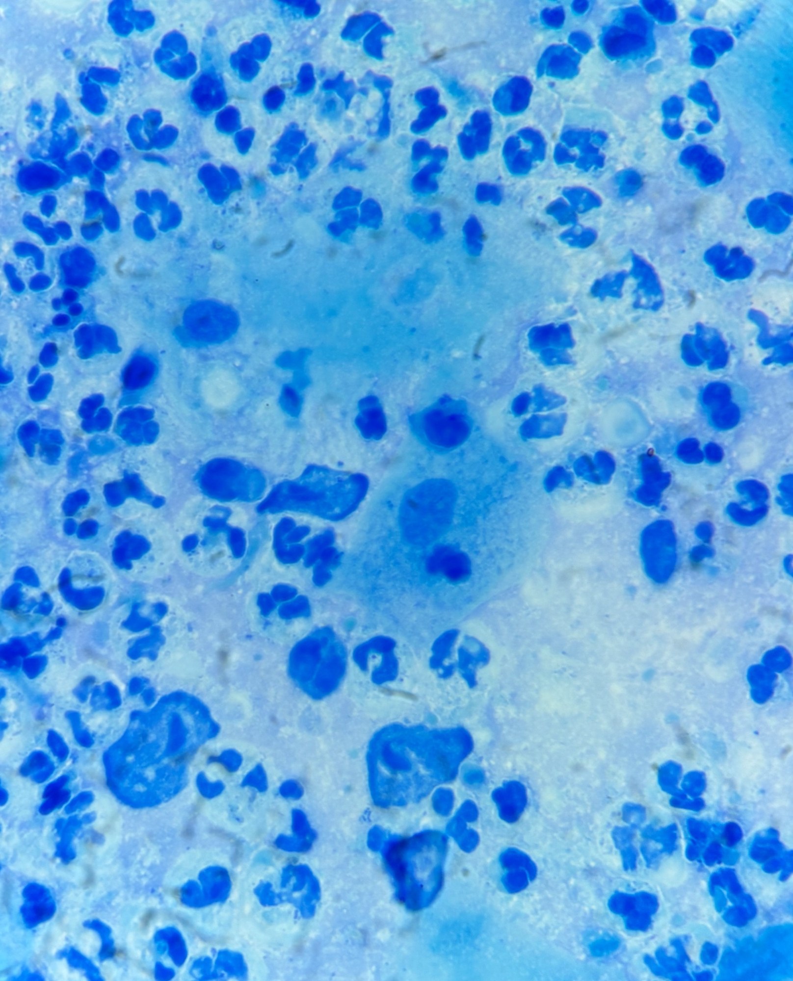
“Beauties” in a female smear. Locus - C
Publish anonymously.
In locus V, it was “classic” - delicate cytoplasm, excellent intense nuclear staining. Unfortunately, I was unable to photograph it.

Microscopic appearance of Trichomonas vaginalis in a stained male flora smear: leukocyte reaction, epithelial cells, and PCR confirmation.

Morphology of Trichomonas vaginalis in stained urogenital smears: identification of protozoa, leukocyte reaction, and bacterial flora during microscopy.

Microscopic image of a mixed infection: Candida pseudohyphae and trichomonads in a flora smear, indicating vaginal candidiasis and trichomoniasis.