

In a female smear, trichomonads were found 🔬
🙏 The publication was kindly provided by Anastasia Kuzmina
Detection of Trichomonas vaginalis during microscopic examination of a flora smear. Morphology of protozoa against the background of a pronounced neutrophilic leukocyte reaction.
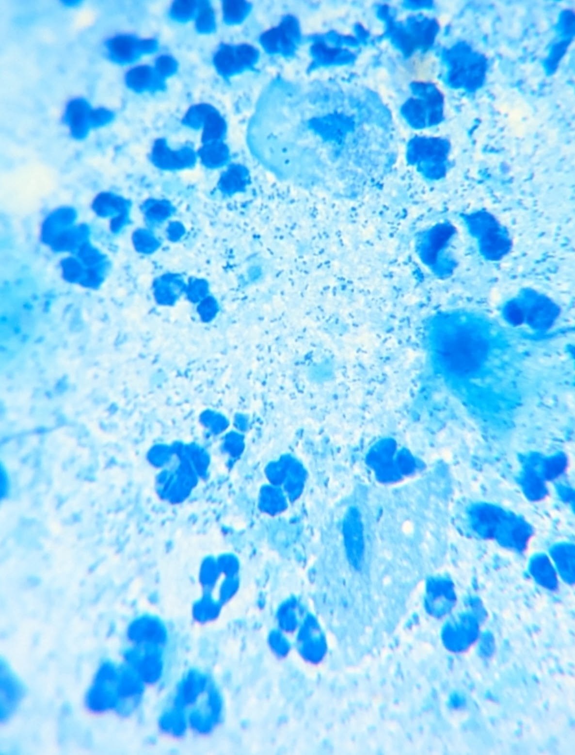
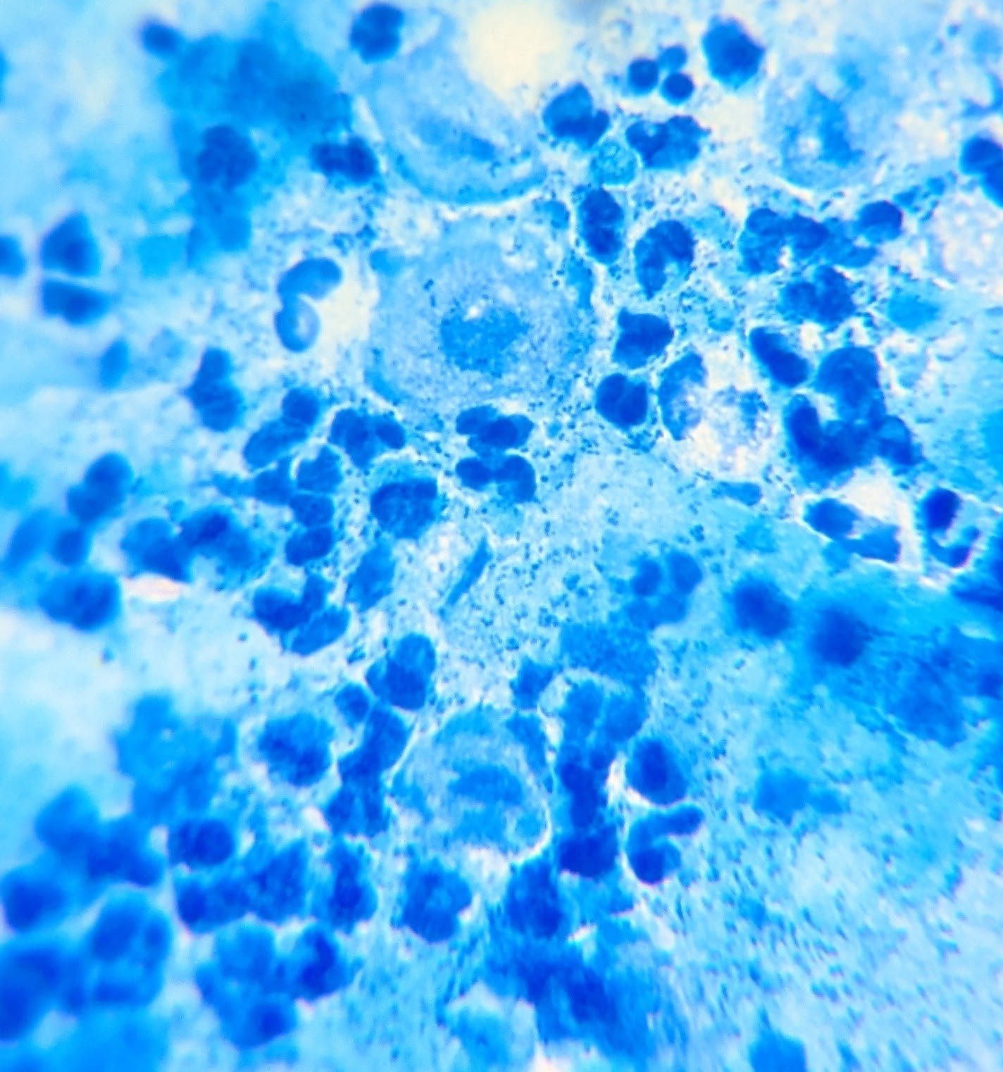
In a female smear, trichomonads were found 🔬
🙏 The publication was kindly provided by Anastasia Kuzmina

Features of detecting Trichomonas vaginalis in smear microscopy: differential diagnosis with leukocytes amidst pronounced inflammation.

Microscopic view of a urogenital smear: identifying Trichomonas vaginalis trophozoites among squamous epithelial cells and abundant microflora.

Microscopic appearance of Trichomonas vaginalis in a stained male flora smear: leukocyte reaction, epithelial cells, and PCR confirmation.