

Strawberry trichomes under the microscope ×10 – do not confuse them with nematodes!
🙏 Photos kindly provided by clinical laboratory diagnostics physician Olga Parfentieva
Microscopy of strawberry trichomes at ×10 magnification: morphological features of plant hairs and differential diagnosis with nematode larvae.
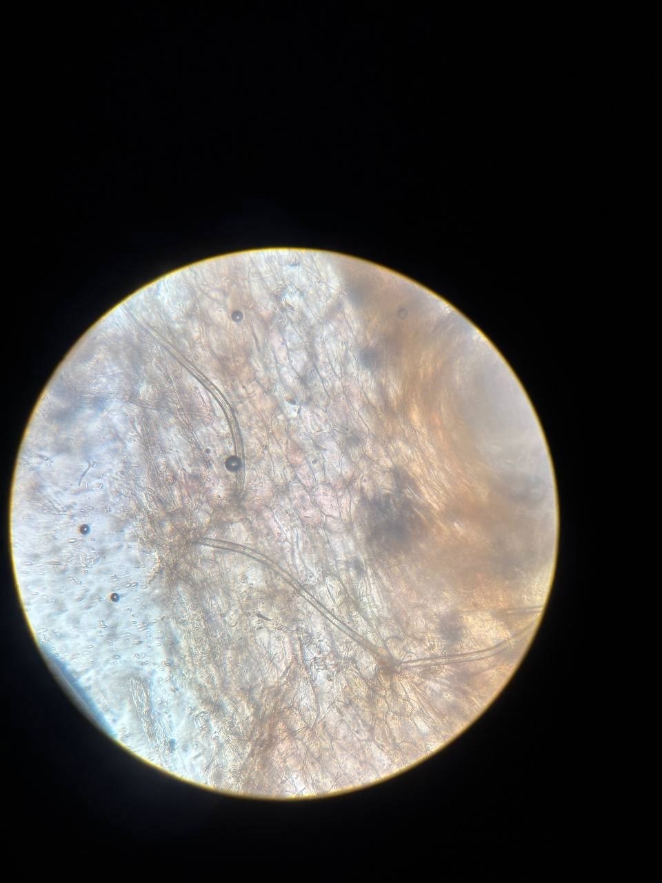
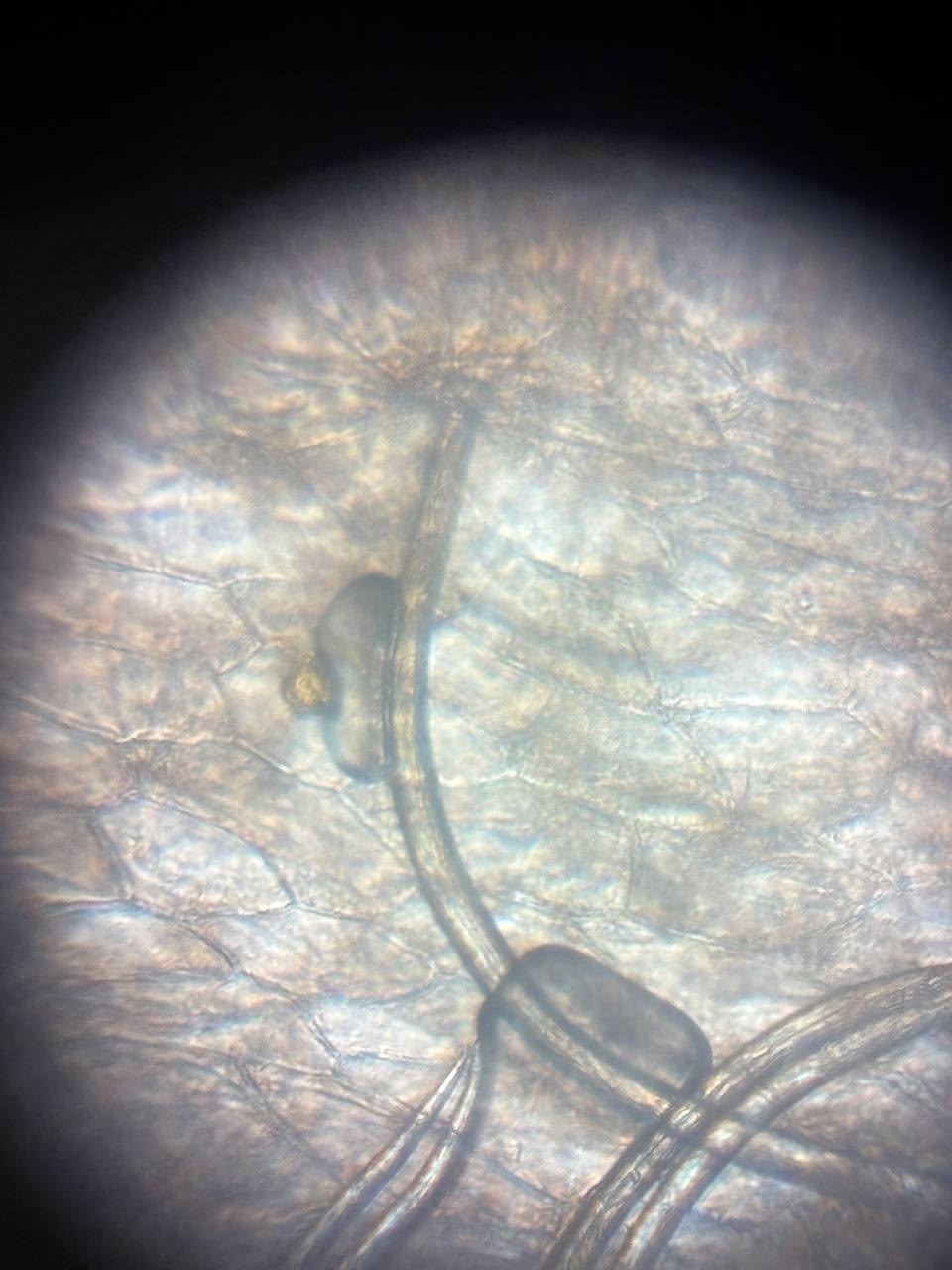
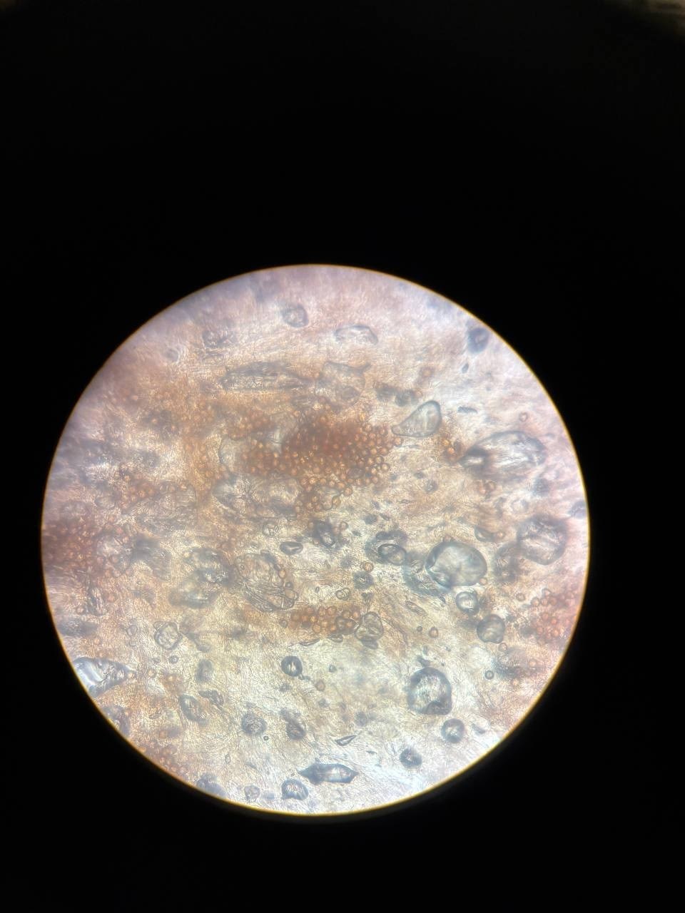
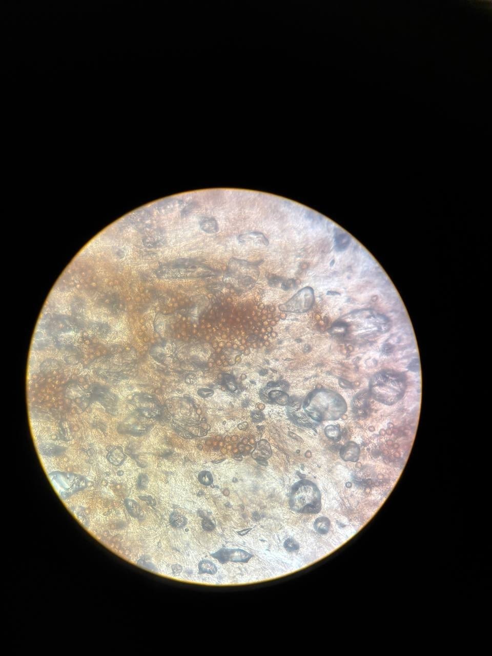
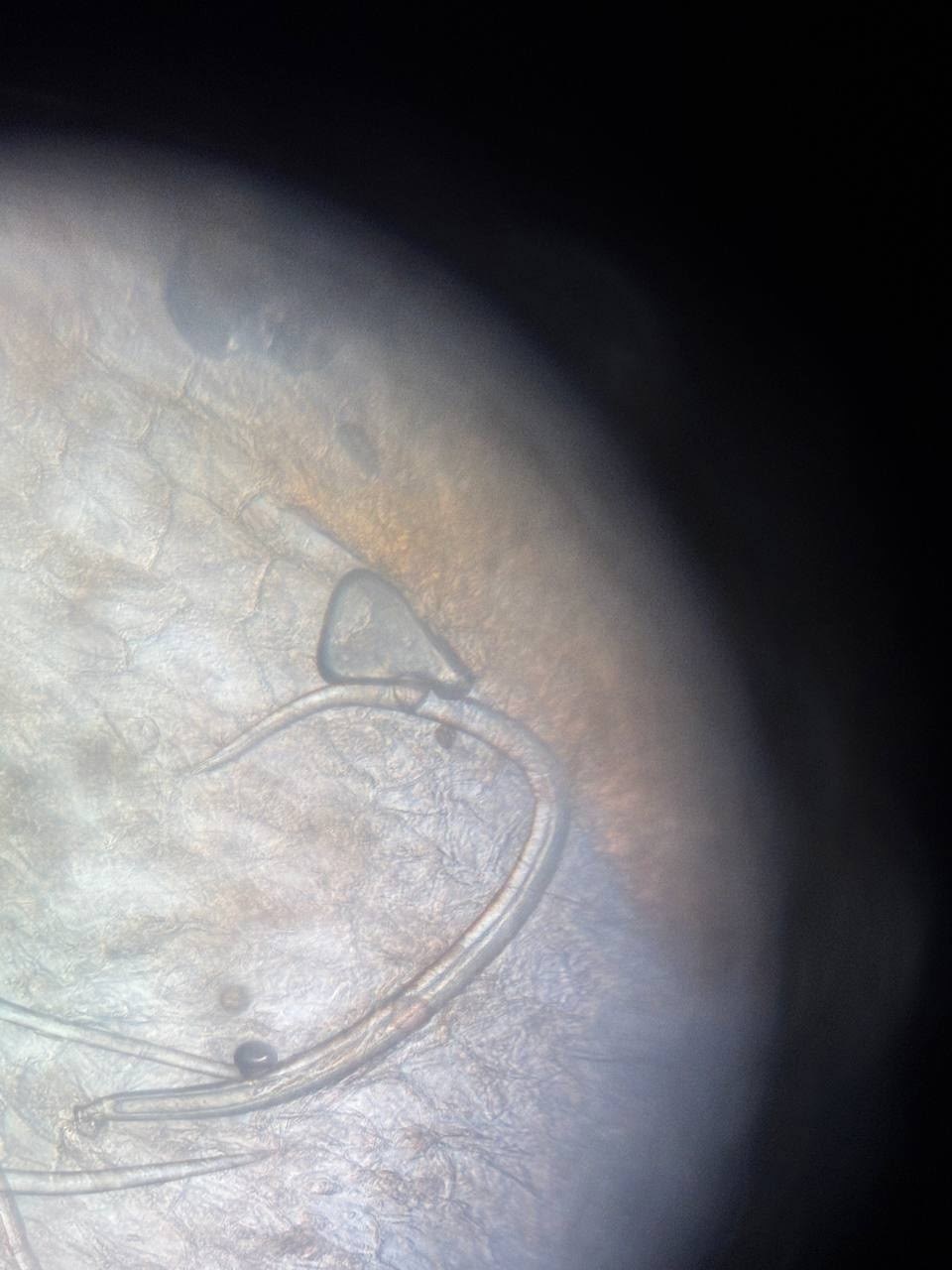
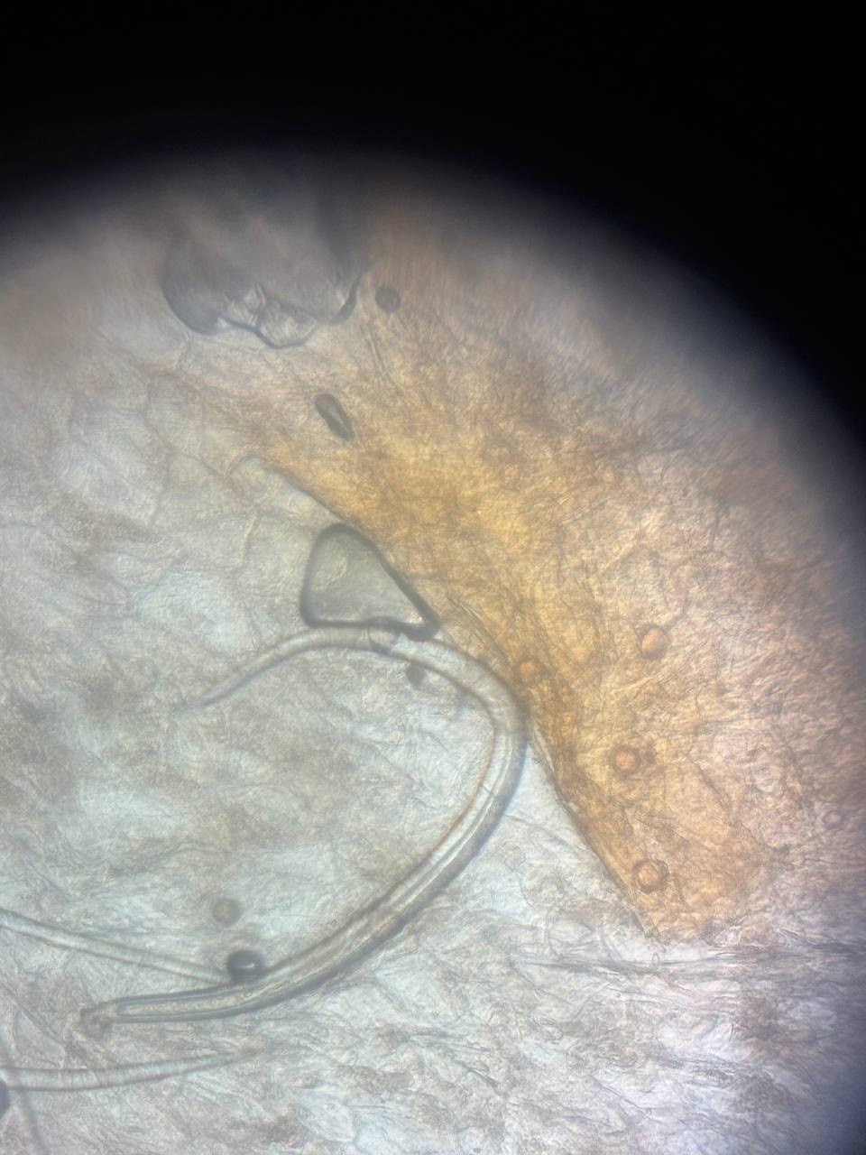
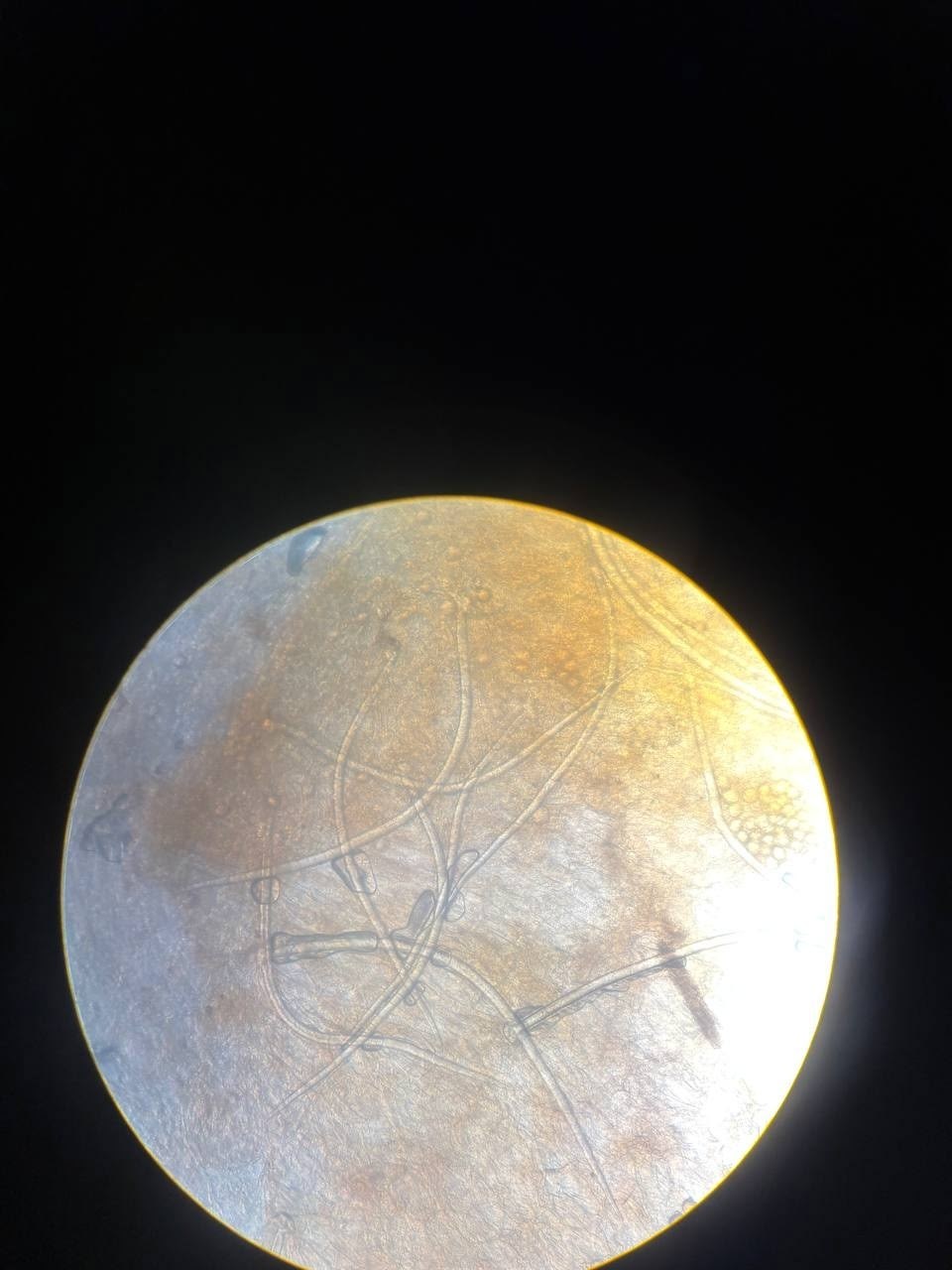
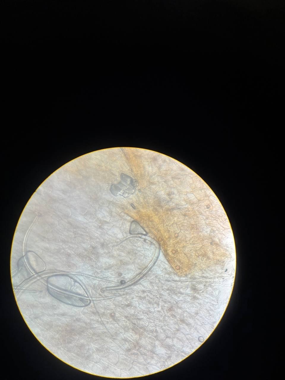
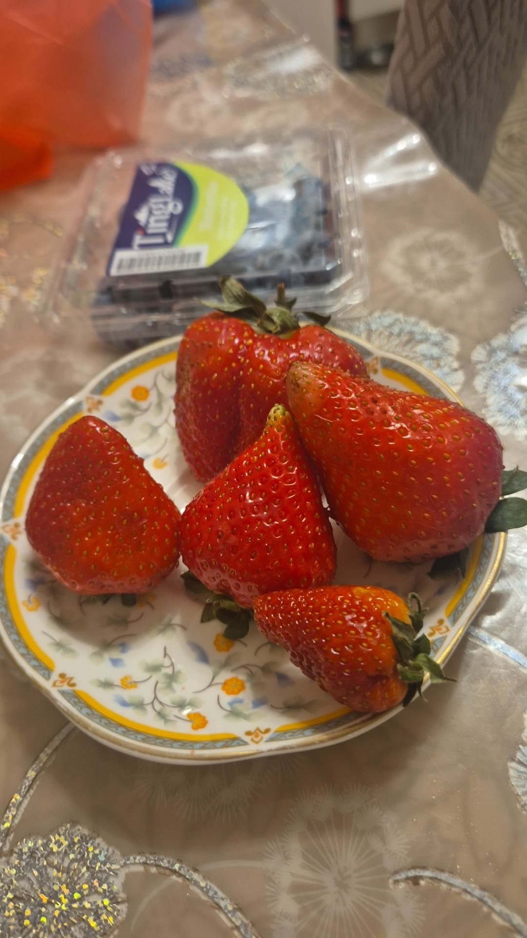
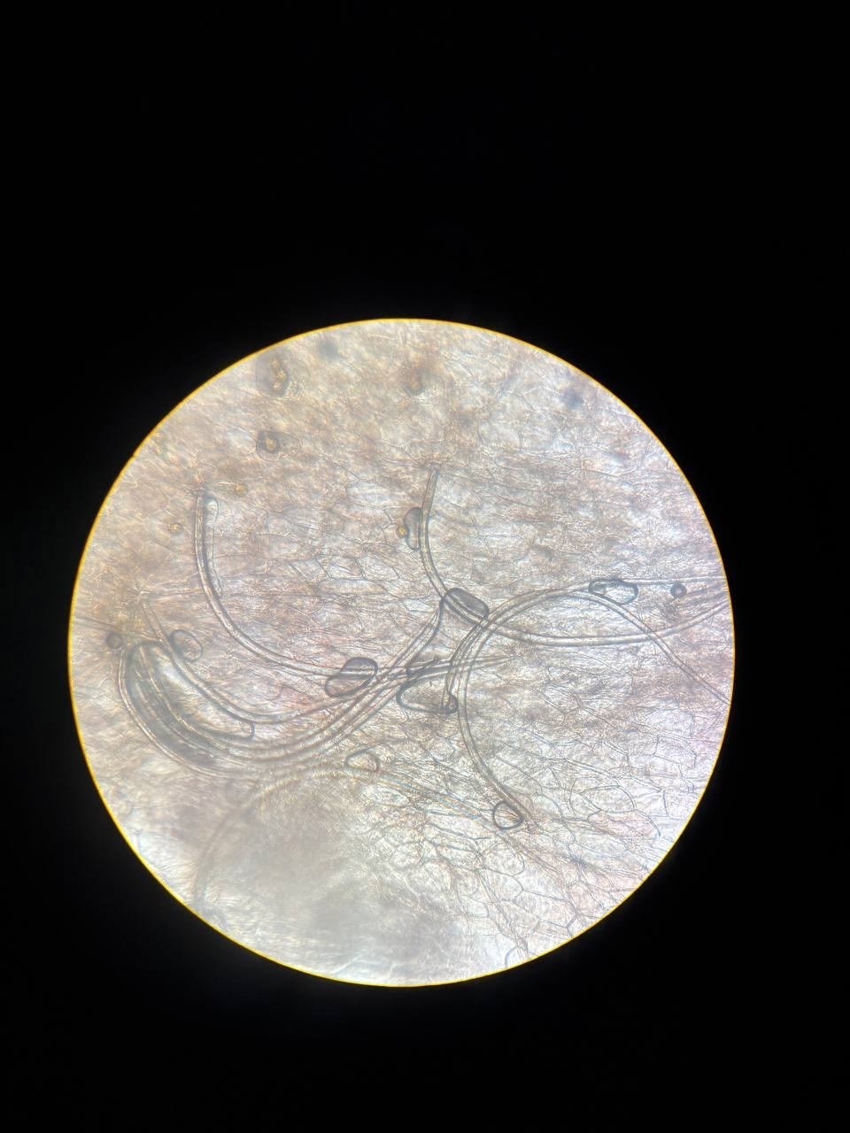
Strawberry trichomes under the microscope ×10 – do not confuse them with nematodes!
🙏 Photos kindly provided by clinical laboratory diagnostics physician Olga Parfentieva

An analysis of native fecal smear microscopy using the KEYU F40 analyzer, focusing on the differential diagnosis of parasitic eggs and plant artifacts.

A clinical case illustrating the differential diagnosis of helminth eggs and plant artifacts during stool microscopy. Key criteria for parasite identification.

Microscopic features of Eucoleus (Thominx) aerophilus lung capillaria eggs: polar plugs, thin shell, and asymmetric shape. Learn more.