

🔬Entamoeba histolytica trophozoites. ✅Dysentery amoeba trophozoite in an iodine-stained wet mount stool sample (bar scale - 12 µm)
Source: Barrios R., Haque A. K. (ed.). Parasitic diseases of the lungs. – Springer, 2013.
Microscopic identification of Entamoeba histolytica trophozoites in a wet mount stool sample stained with Lugol's iodine solution.
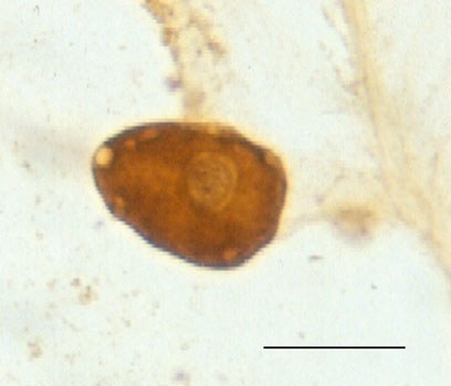
🔬Entamoeba histolytica trophozoites. ✅Dysentery amoeba trophozoite in an iodine-stained wet mount stool sample (bar scale - 12 µm)
Source: Barrios R., Haque A. K. (ed.). Parasitic diseases of the lungs. – Springer, 2013.

Morphological features of Entamoeba histolytica and Entamoeba dispar cysts in stool microscopy. Routes of infection and prevention measures for intestinal amebiasis.

A diagnostic microscopy exercise: identify parasitic objects among elements and artifacts in the field of view. Visual recognition training.

An interactive microscopy challenge in parasitology: identify and locate a parasite object among artifacts and coprogram elements within a limited time.