

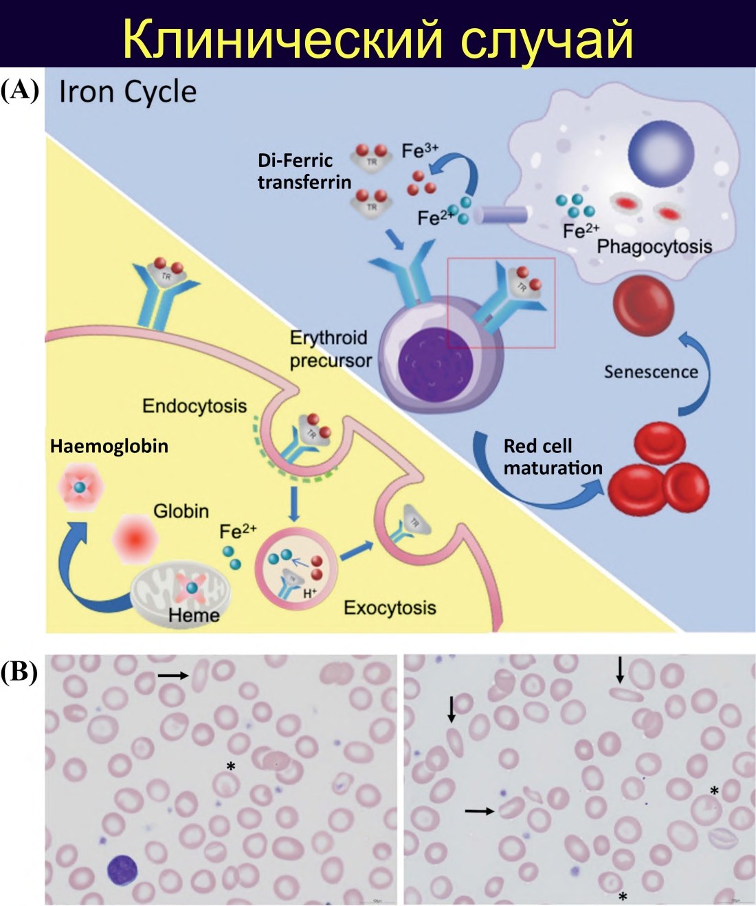
Iron Deficiency Anemia A 45-year-old female patient, previously healthy. For 3 months, she has noted increasing dyspnea on exertion: she used to easily run at least 5 km, but now she has to stop several times. At rest, she feels a rapid and “strong” heartbeat. Menstruation is regular, and according to the patient, “normal in volume.” Stool is without impurities, appetite is preserved. She does not smoke or consume alcohol.
✅ Laboratory parameters (initial blood test and iron metabolism) Hemoglobin: 69 g/L (↓) Mean corpuscular volume: 63.6 fL (microcytosis) Mean corpuscular hemoglobin: 17.7 pg (↓) Mean corpuscular hemoglobin concentration: 27.8 g/dL (↓) White blood cells: 5.07 × 10⁹/L (normal) Platelets: 479 × 10⁹/L (reactive thrombocytosis)
Serum iron: 1.7 µmol/L (markedly ↓) Total iron-binding capacity: 87.4 µmol/L (↑) Transferrin saturation: 2 % (↓)
Laboratory interpretation: a typical profile of severe iron deficiency with compensatory increase in transferrin synthesis and extremely low saturation.
✅ Peripheral blood smear Hypochromic microcytic anemia with marked anisocytosis and poikilocytosis:
- small, pale red blood cells with an enlarged central pallor
- elliptocytes
- target cells
- thin, elongated elliptocytes — so-called “pencil” cells
This morphological profile is well consistent with severe iron deficiency anemia and reactive thrombocytosis.
✅ Diagnostic conclusion The totality of data:
- severe hypochromic microcytic anemia
- low serum iron
- elevated total iron-binding capacity
- low transferrin saturation
- characteristic smear morphology
— allows for a confident conclusion: severe iron deficiency anemia.
For complete diagnosis and monitoring, from a laboratory perspective, it is advisable to:
- determine ferritin (with the caveat that it is an acute phase protein)
- assess inflammatory markers (e.g., C-reactive protein) to more accurately interpret ferritin
- if necessary — dynamic observation of red blood cell indices and platelets during therapy
✅ Search for the cause of iron deficiency (clinical and laboratory context) In a woman of reproductive age, the most likely source of chronic blood loss is uterine bleeding, even with “normal” menstruation according to the patient. Recommended:
- gynecological examination with pelvic ultrasound (search for uterine fibroids, endometrial hyperplasia, etc.)
- gastroenterological examination if gastrointestinal symptoms are present or if the source of blood loss is unclear (gastritis, peptic ulcer disease, tumors, angiodysplasia, etc.)
- in rare cases — exclusion of paroxysmal nocturnal hemoglobinuria if episodes of dark urine and laboratory signs of intravascular hemolysis are present
For a laboratory hematologist, it is important in the conclusion not only to state iron deficiency anemia but also to emphasize the need to search for chronic blood loss, especially in severe anemia.
✅ Differential diagnosis (important when describing the smear and conclusion) Main alternatives for hypochromic microcytic anemia in adults:
- thalassemia minor (or another mild form of thalassemia) — typically marked microcytosis with relatively moderate anemia; family history, possible increase in hemoglobin fractions according to hemoglobin spectrum analysis
- anemia of chronic disease with microcytosis — usually reduced or normal total iron-binding capacity and normal or elevated ferritin
Reflecting characteristic morphological features and iron metabolism parameters in the laboratory report helps the clinician reach the correct diagnosis faster.
✅ Treatment (in the context of laboratory support) Main approach:
- oral iron preparations (most often with ferrous salts), usually on an empty stomach, for at least 3–6 months to replenish stores
- in severe anemia, malabsorption, or intolerance to oral preparations — intravenous iron preparations
Laboratory role:
- monitoring dynamics of hemoglobin, red blood cell indices, platelets
- assessing the trend of recovery of parameters 2–4 weeks after the start of therapy
- with intravenous iron administration — determination of ferritin and, if necessary, iron metabolism parameters to avoid iron overload
This clinical case well illustrates how indicative the combination is of:
- marked decrease in hemoglobin,
- characteristic microcytic hypochromic morphology,
- shift in iron metabolism parameters —
for rapid and accurate laboratory diagnosis of iron deficiency anemia and for forming a clear algorithm for further cause-finding for the clinician. Source: Anskar Y.H. Leung - Haematology and the Asian Patient 51 Clinical Cases - 2024


