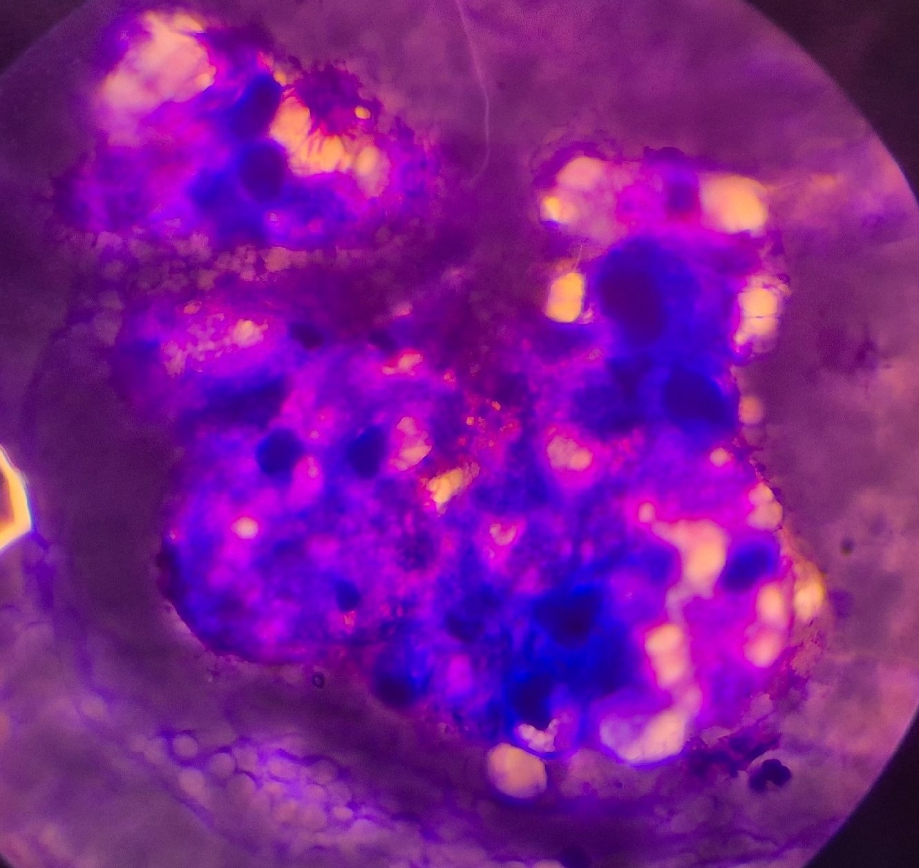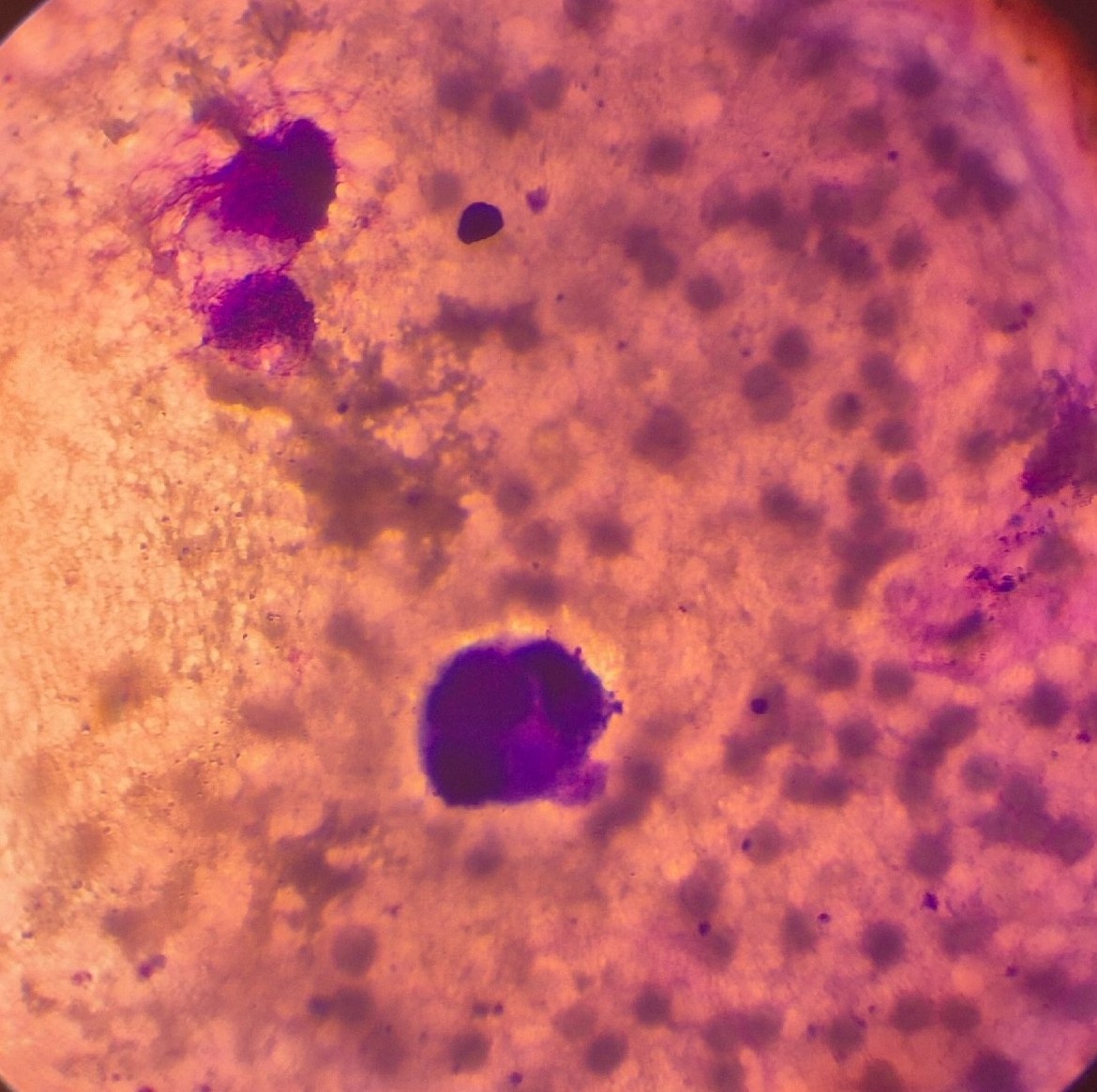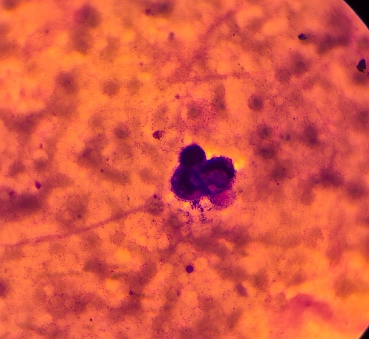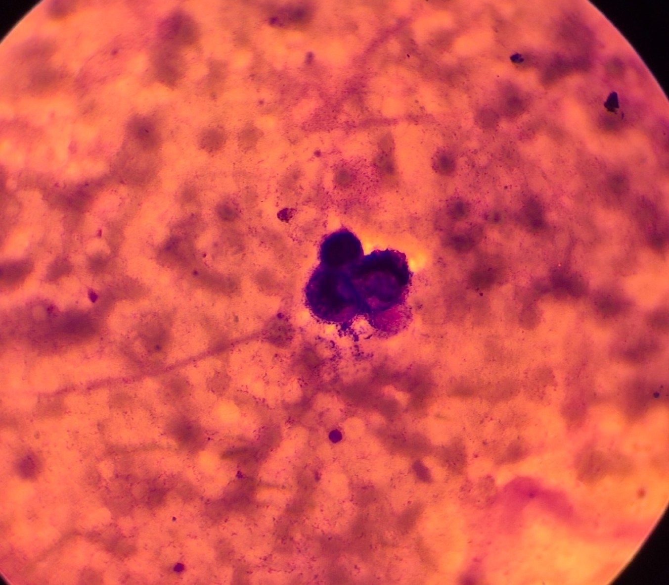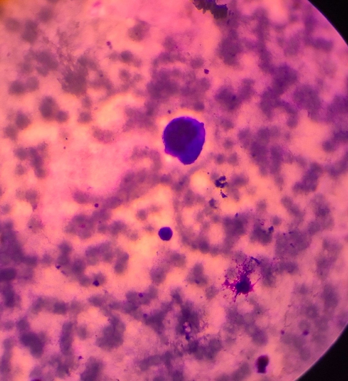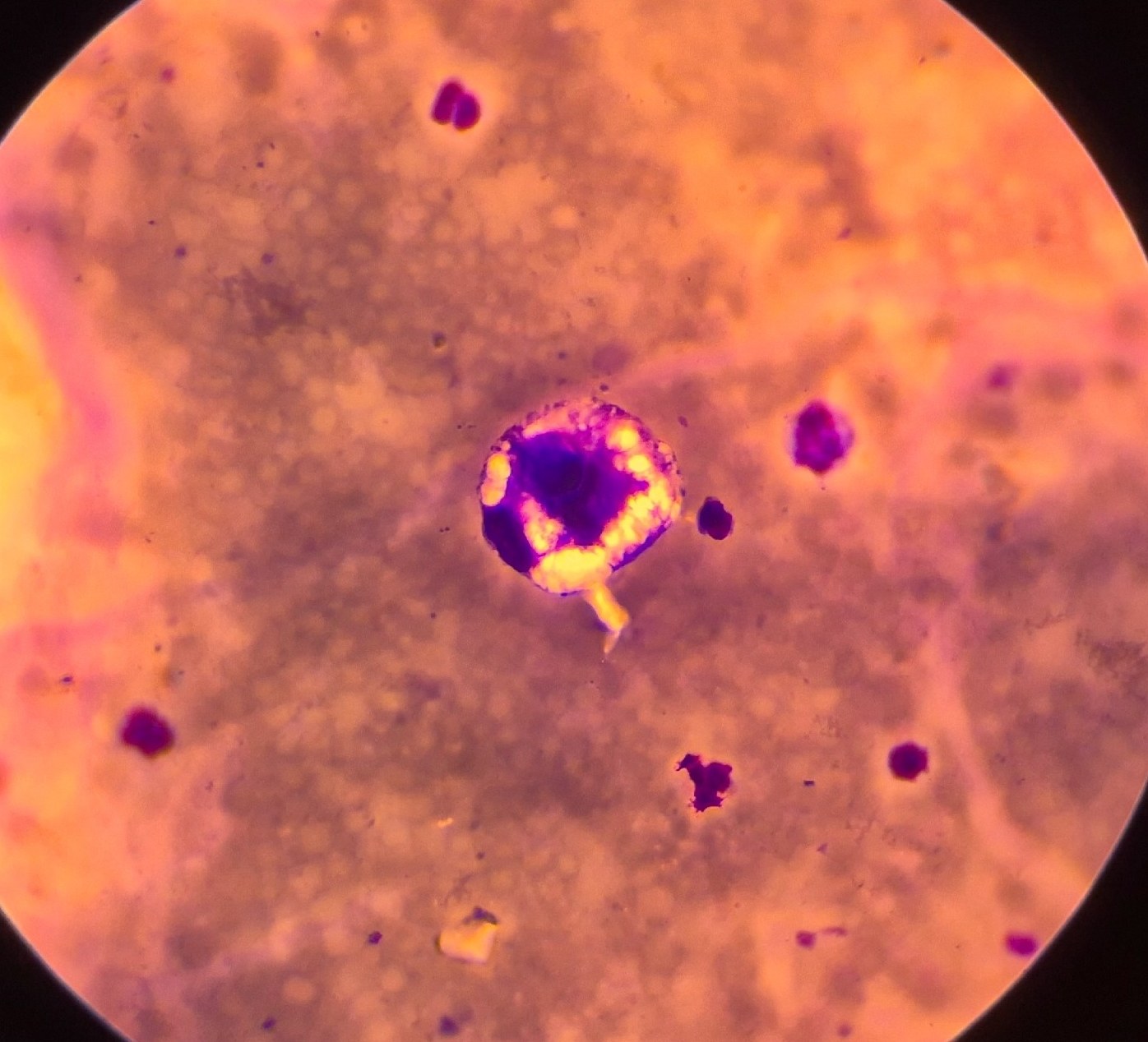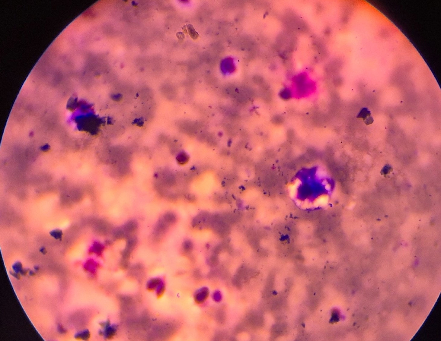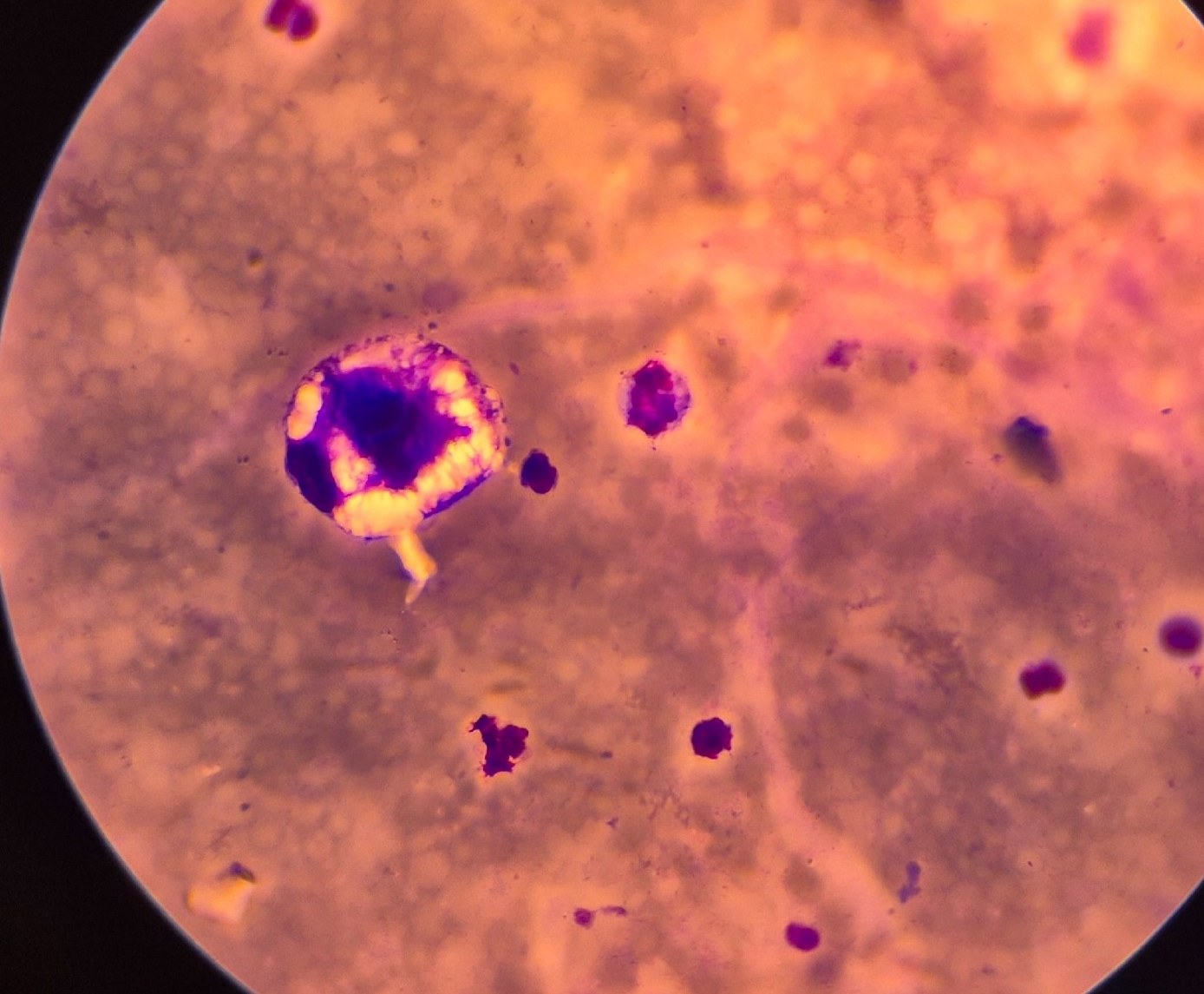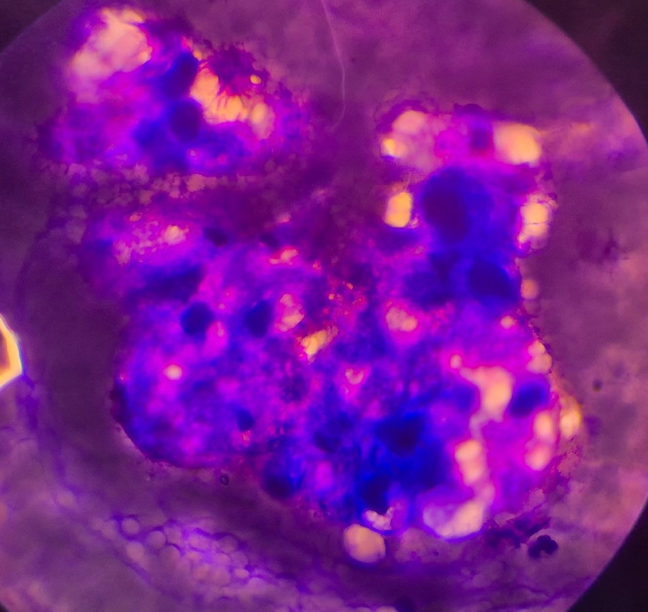
A 56-year-old male patient presented with complaints of hematuria. Reddish-brown urine was delivered to the laboratory. Microscopy of the sediment revealed an abundance of erythrocytes, conglomerates of altered epithelium with atypical, elongated “tailed” cells. It was decided to additionally stain the sediment.
The patient was referred for ultrasound, which revealed a mass in the bladder. Subsequent cystoscopy, biopsy, and histological examination confirmed the diagnosis: urothelial carcinoma.
Fixative: May-Grünwald, Romanowsky stain
🙏 Case kindly provided by clinical laboratory diagnostics physician Olga Parfentieva
Photos