

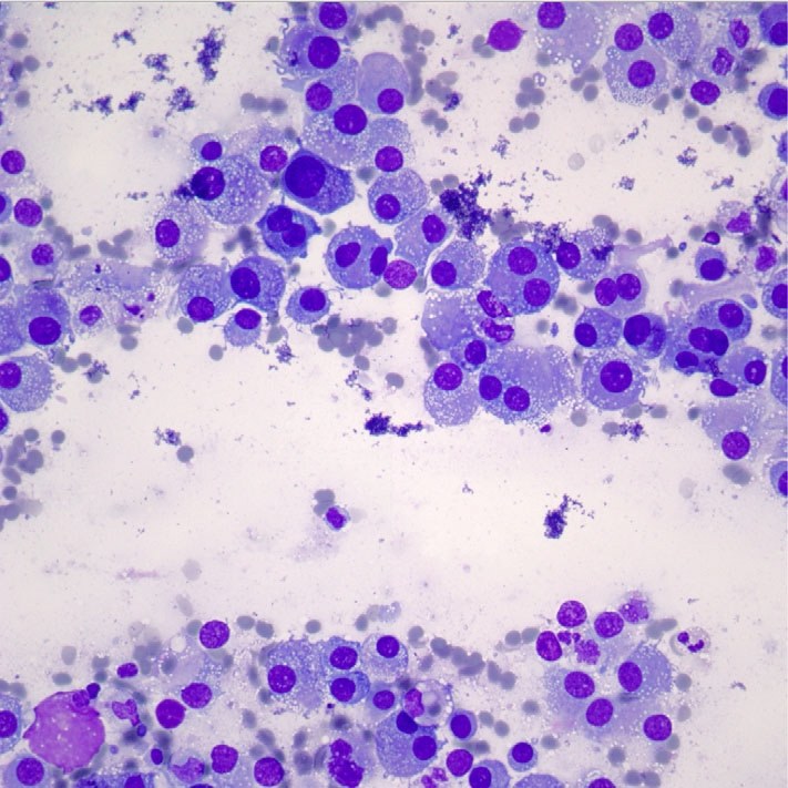
🔬 Cytology of pleural effusion. The journal Diagnostic Cytopathology described a unique case of urothelial carcinoma detection in pleural effusion. Smears revealed small to medium-sized tumor cells, solitary and in small groups. Their main feature was prominent intracellular vacuoles, which could have been mistaken for macrophages. 📌 Additional tests Immunohistochemical staining showed: ✅ Nuclear expression of GATA3, CK7, and thrombomodulin ❌ Negative reactions for CD68 and CK20 🔴 Oil-Red staining revealed lipids within the vacuoles.
Source: Saganas C, Hewer E. Lipidised urothelial carcinoma in pleural effusion—A case report. Diagnostic Cytopathology. 2020;1–2.


