

🔬 Vaginal Candidiasis + Trichomoniasis
🙏 Image kindly provided by Laboratory Diagnostics Physician Alena Chirvina, Rostov-on-Don
Microscopic image of a mixed infection: Candida pseudohyphae and trichomonads in a flora smear, indicating vaginal candidiasis and trichomoniasis.
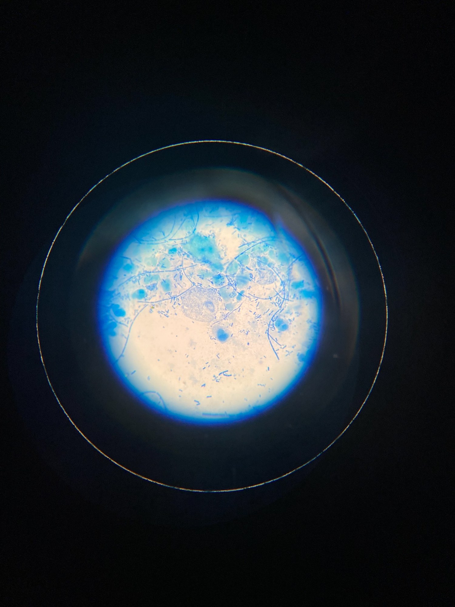
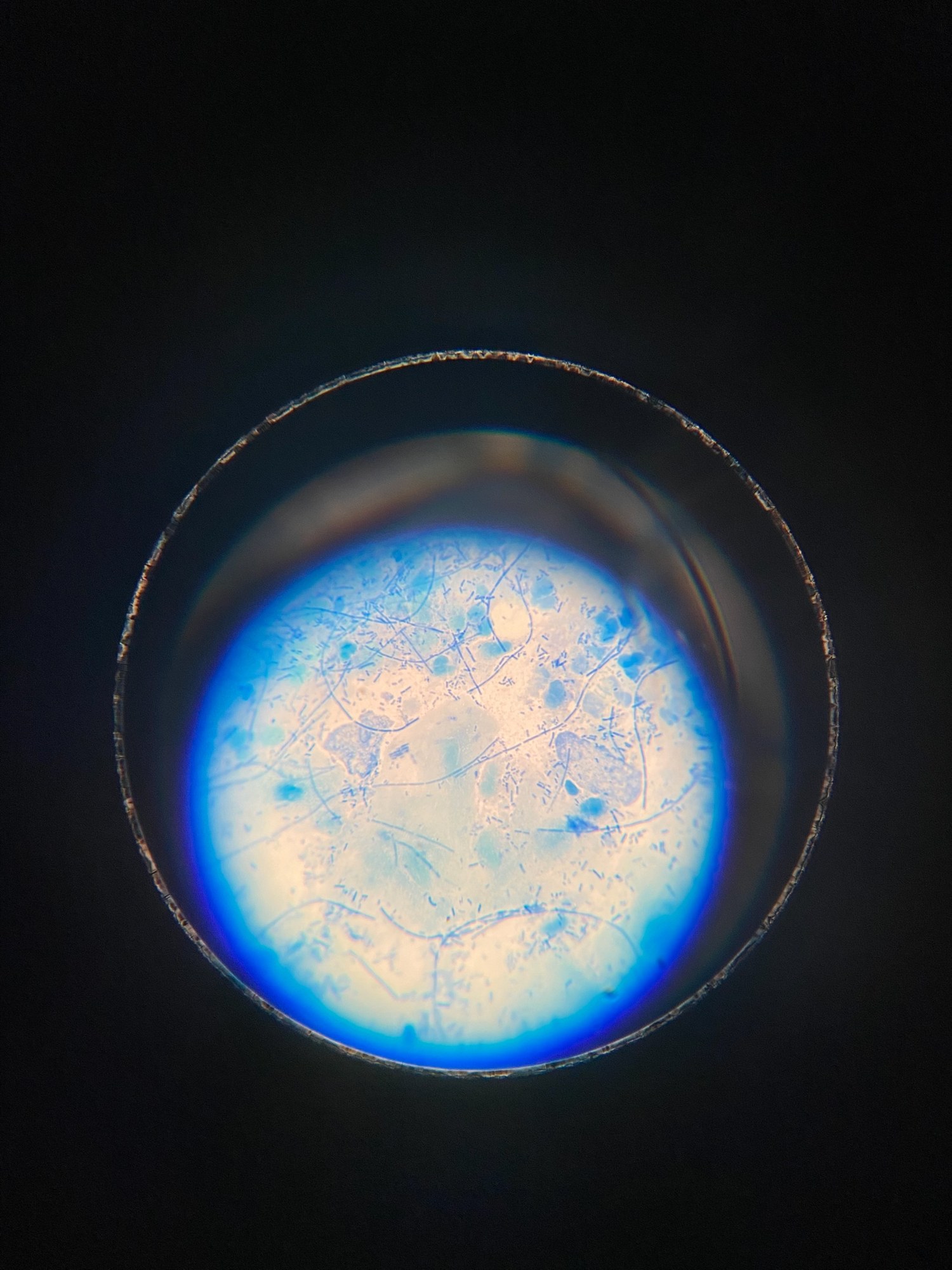
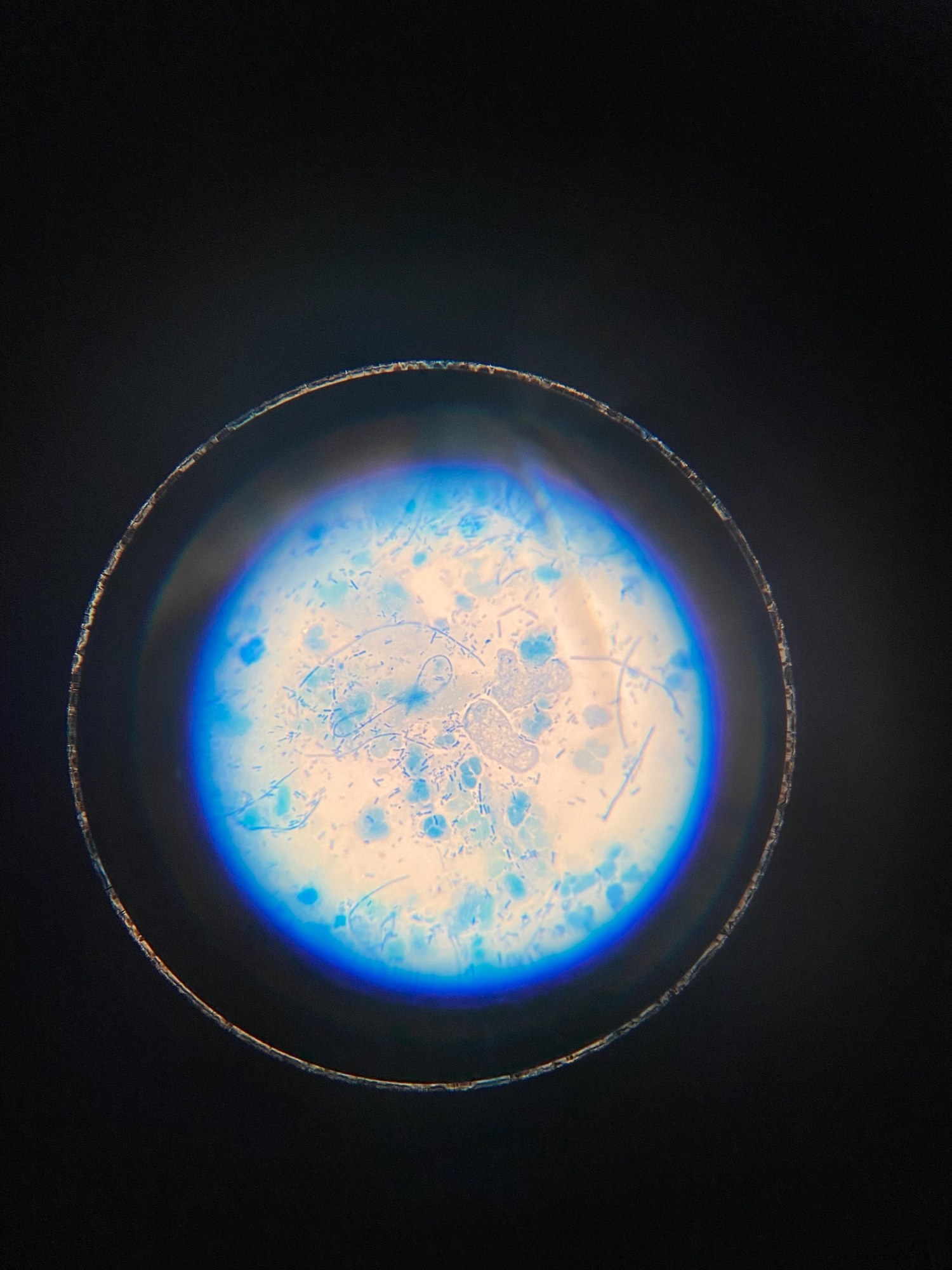
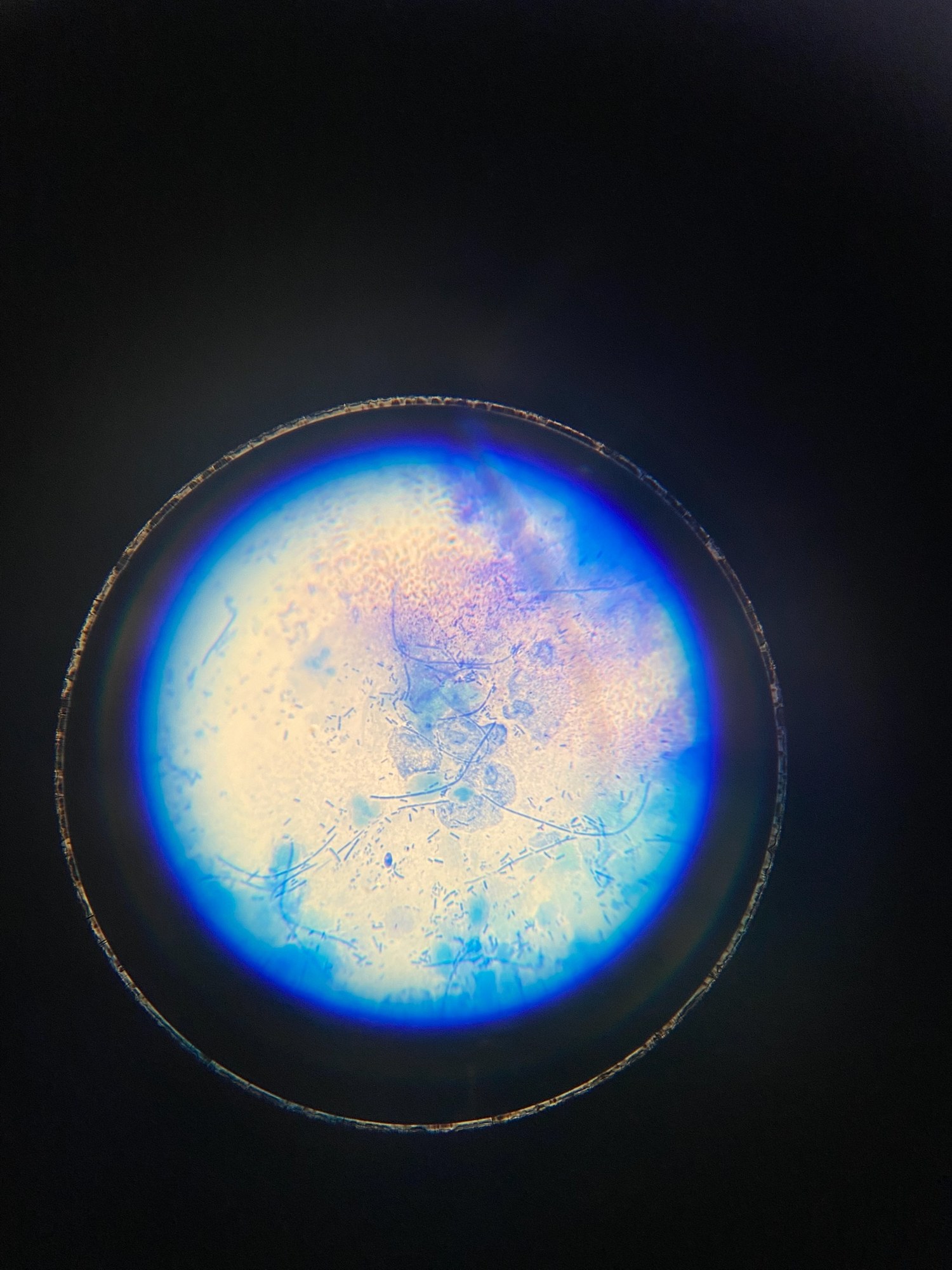
🔬 Vaginal Candidiasis + Trichomoniasis
🙏 Image kindly provided by Laboratory Diagnostics Physician Alena Chirvina, Rostov-on-Don

Microscopic appearance of Trichomonas vaginalis in a stained male flora smear: leukocyte reaction, epithelial cells, and PCR confirmation.

Morphology of Trichomonas vaginalis in stained urogenital smears: identification of protozoa, leukocyte reaction, and bacterial flora during microscopy.

Microscopy of a stained gynecological smear: detection of Trichomonas vaginalis with flagella amidst abundant bacterial flora and leukocytes.