

Testicular tissue, dog (normal). Germ cells: mitotic spindle.
👉Image kindly provided by - Olga Belozerova, clinical pathologist, head of the laboratory at the veterinary clinic “Noah’s Ark”, Kirov.
Cytological image of normal canine testicular tissue: dividing germ cells with visible mitotic spindles. Morphology of spermatogenesis.
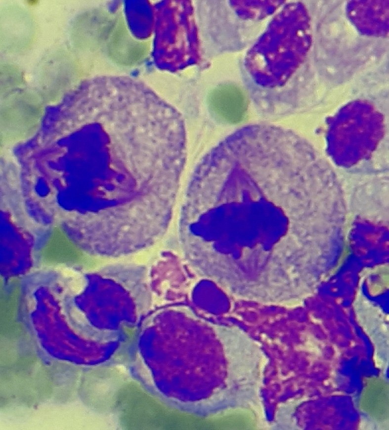
Testicular tissue, dog (normal). Germ cells: mitotic spindle.
👉Image kindly provided by - Olga Belozerova, clinical pathologist, head of the laboratory at the veterinary clinic “Noah’s Ark”, Kirov.

Cytological features of normal mesothelium: cell morphology, round nucleus, basophilic cytoplasm, and characteristic pink border under the microscope.

Key cytological criteria of malignancy: anisocytosis, anisokaryosis, atypical mitoses, nuclear polymorphism, and cell cannibalism in microscopy.

Morphological features of cervical cells with HPV: signs of severe dyskaryosis, hyperchromatic nuclei with folded contours, and scanty cytoplasm.