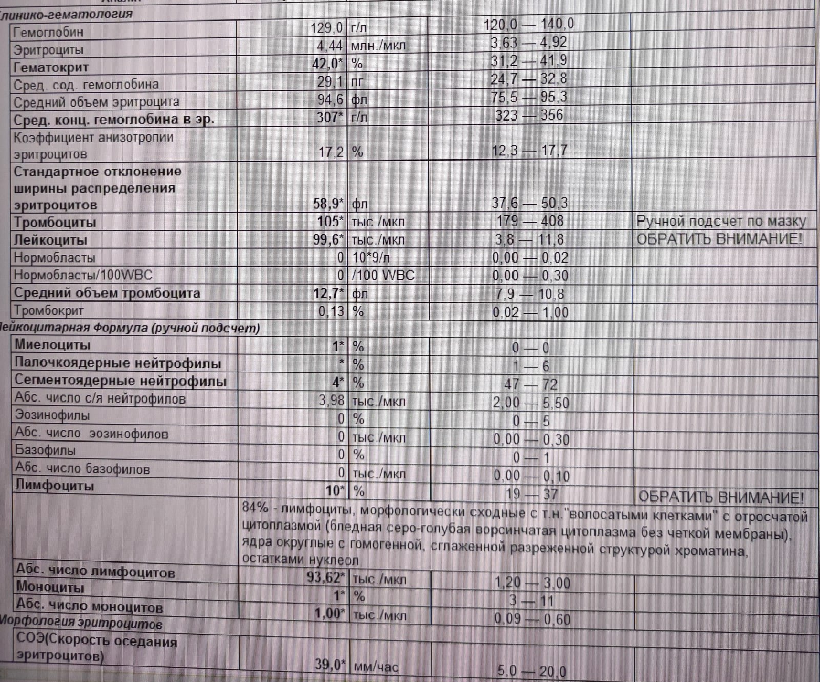
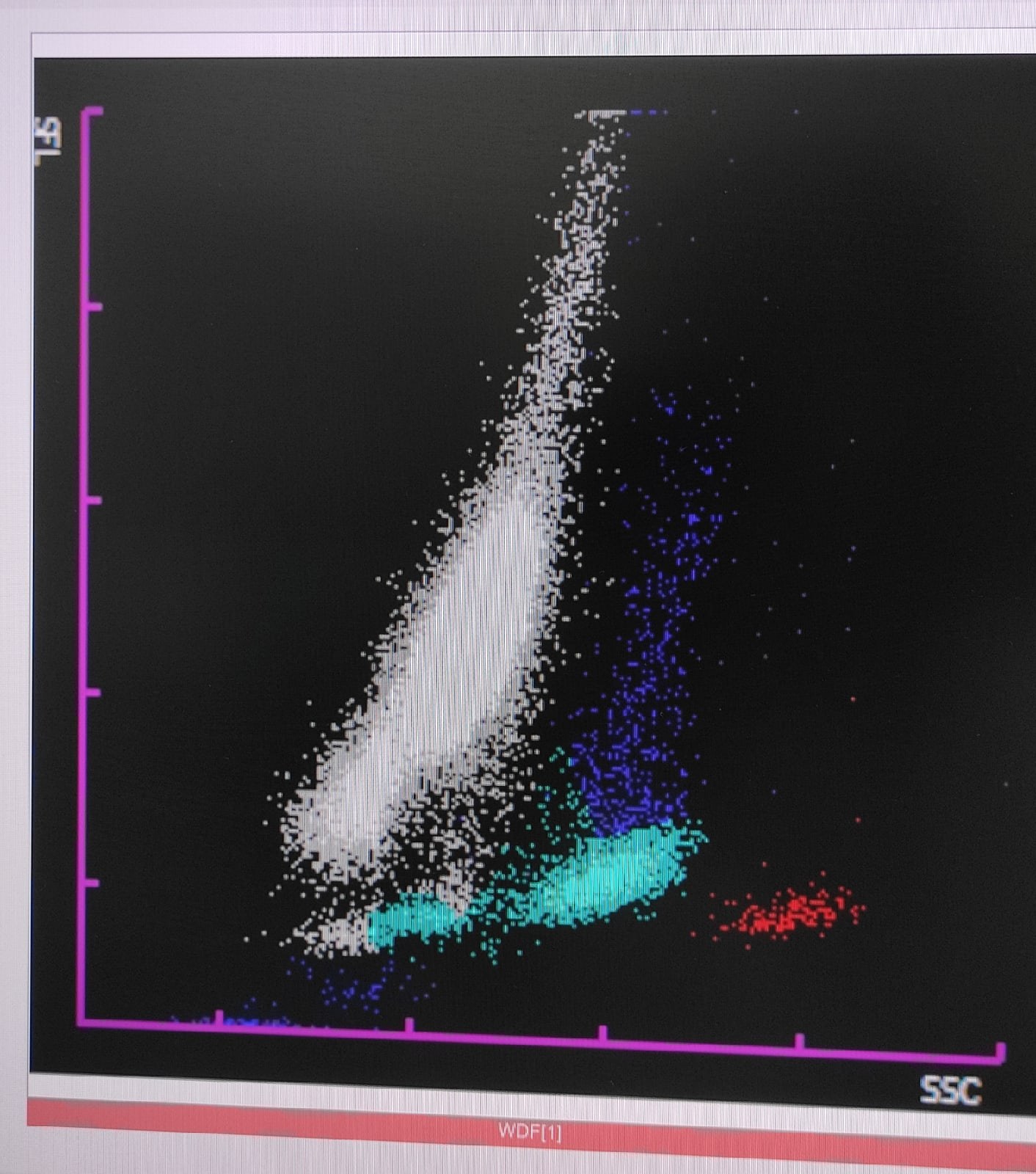
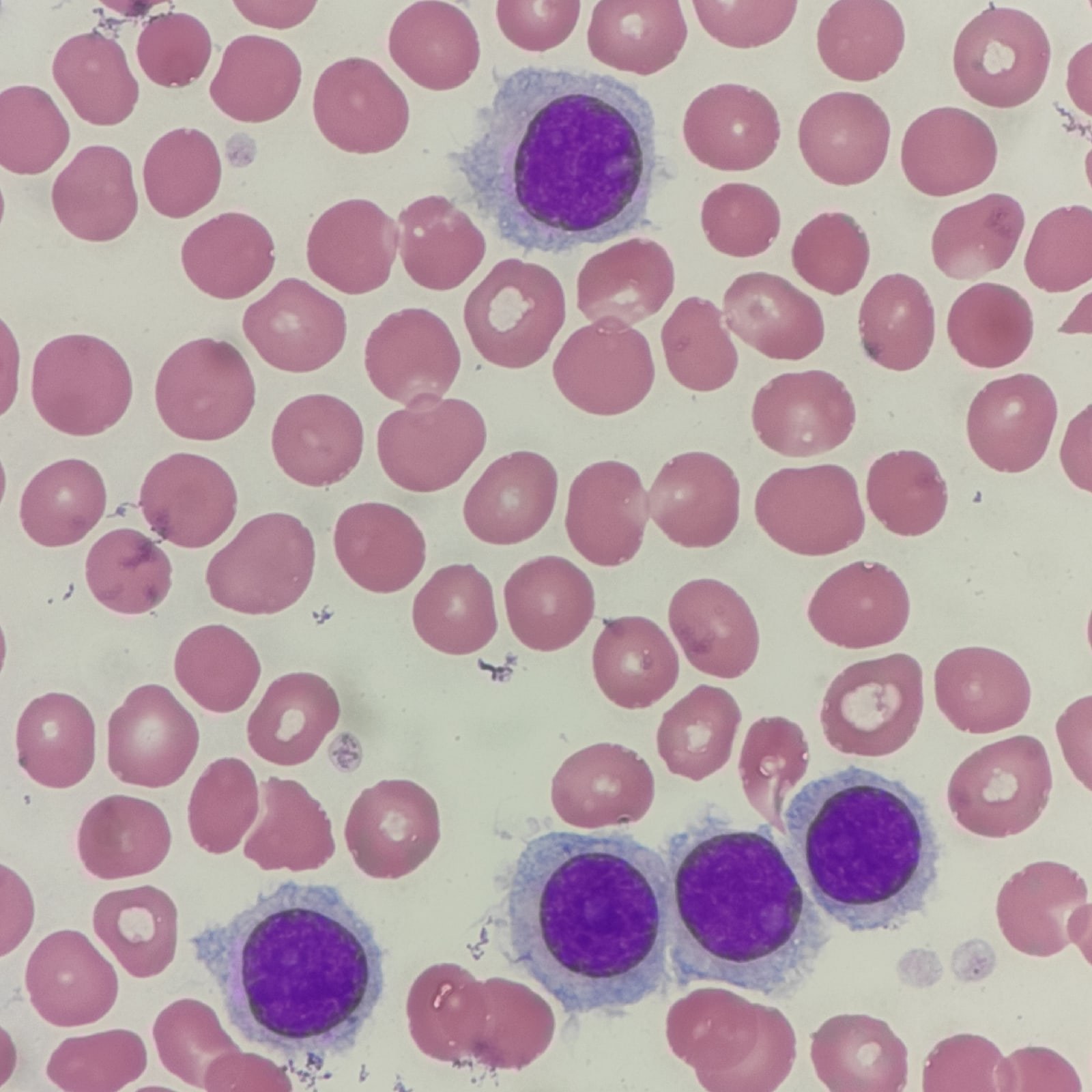
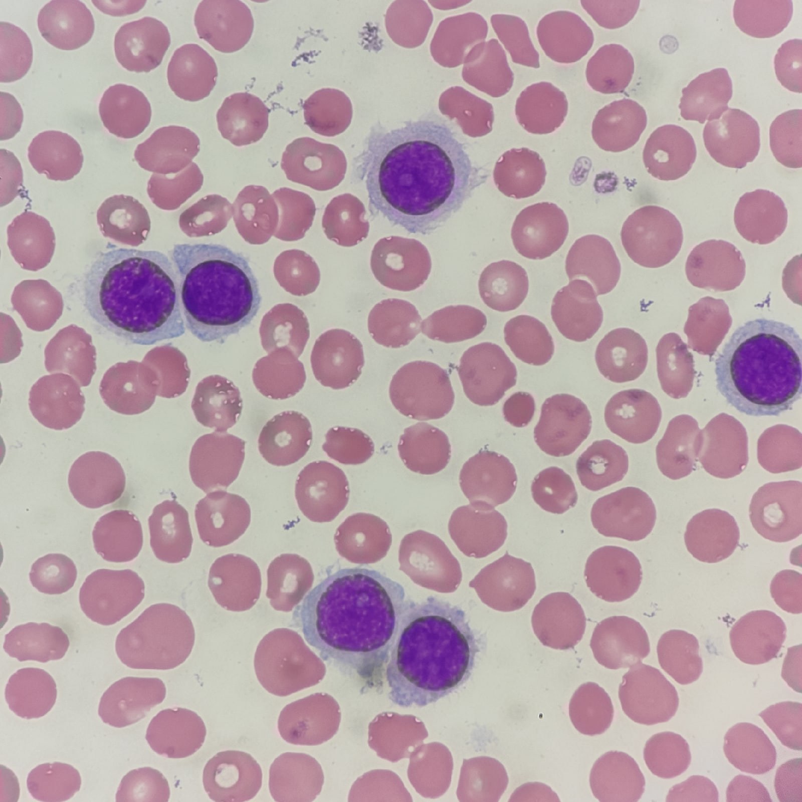
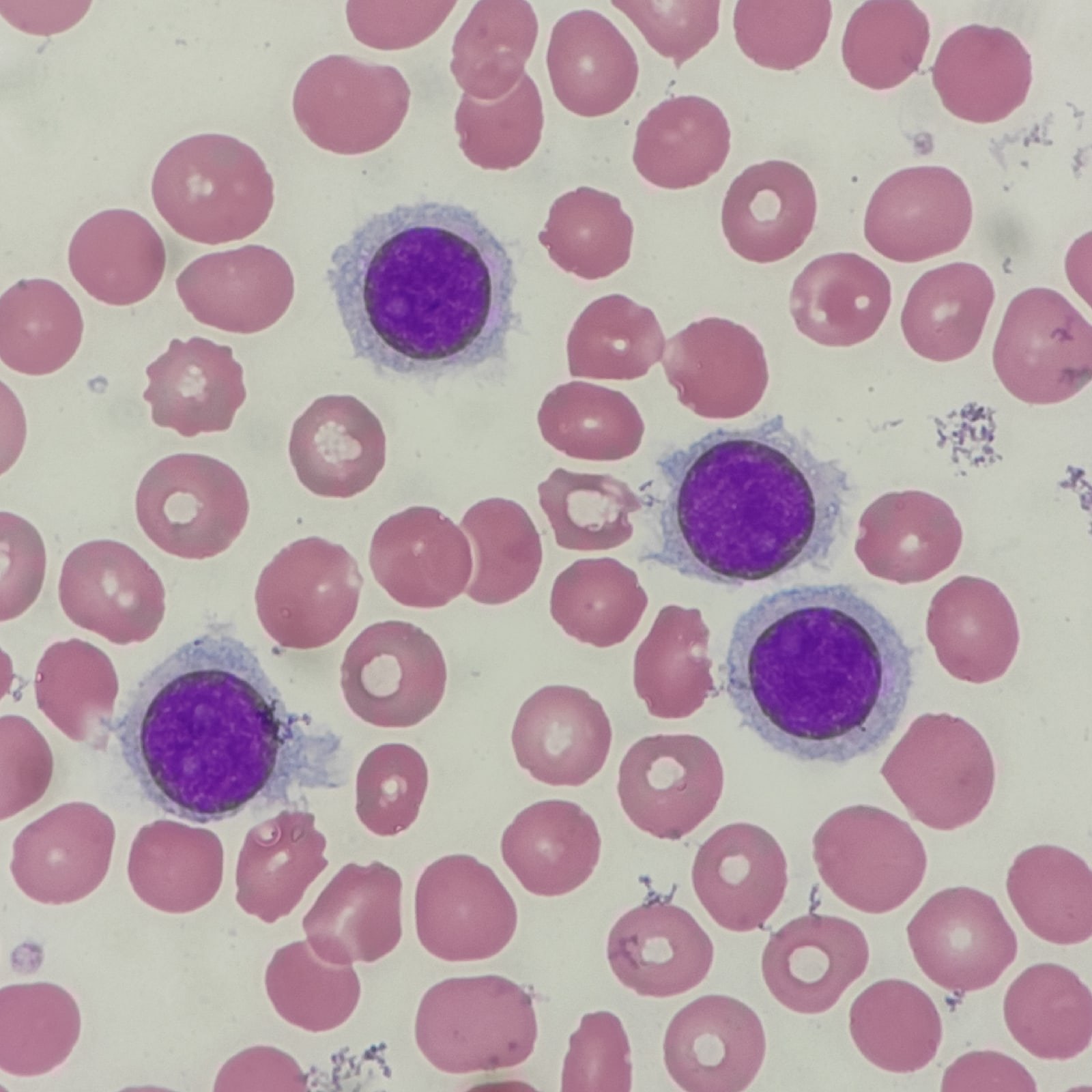
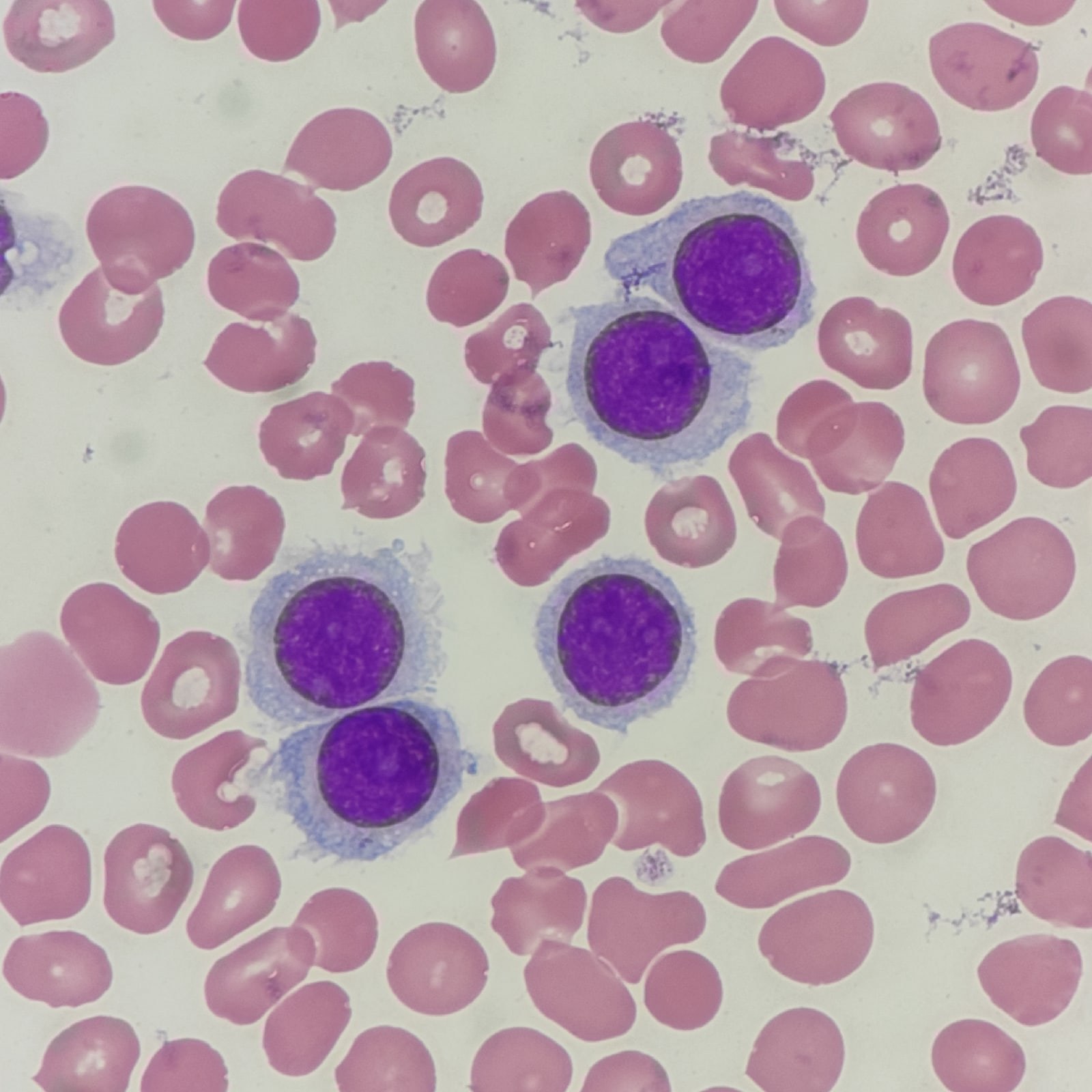
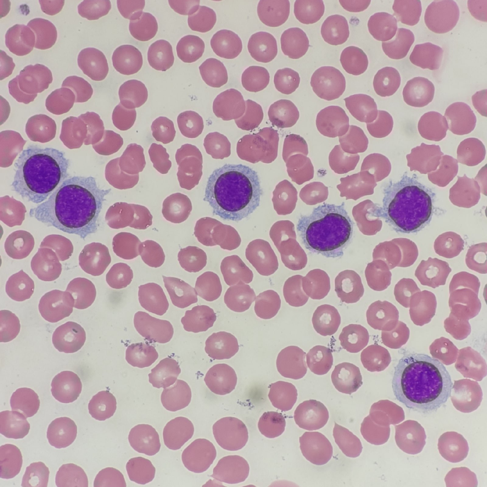
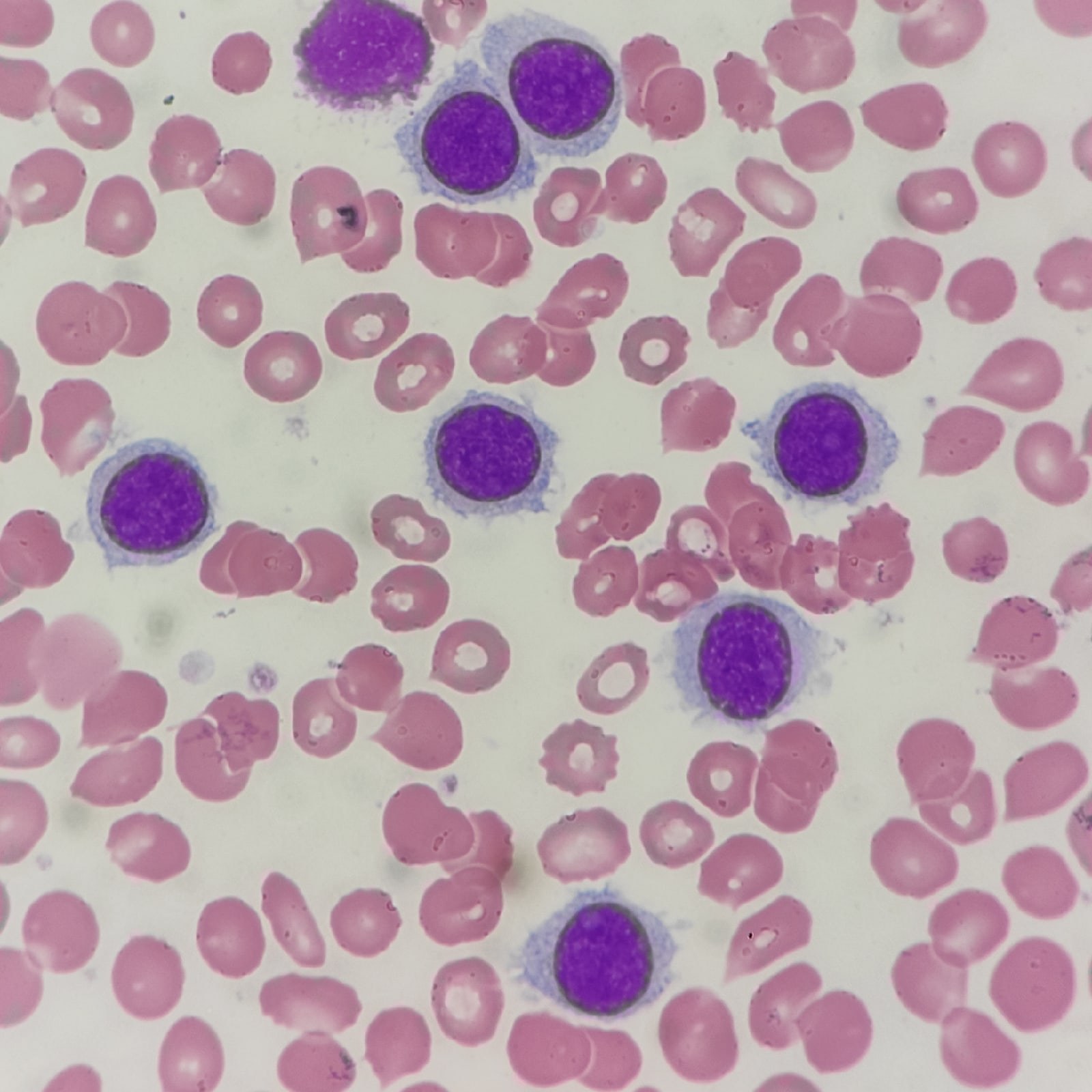
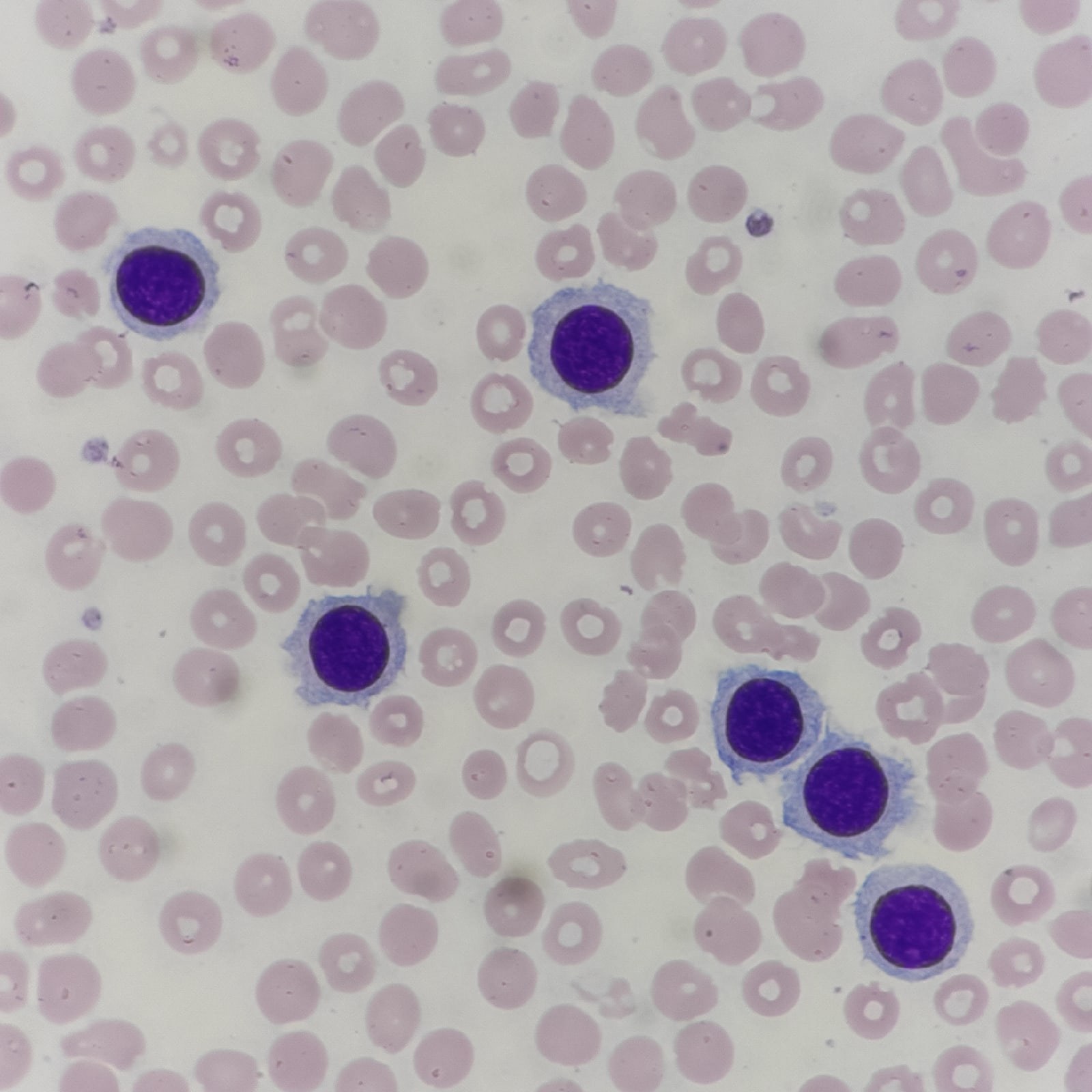
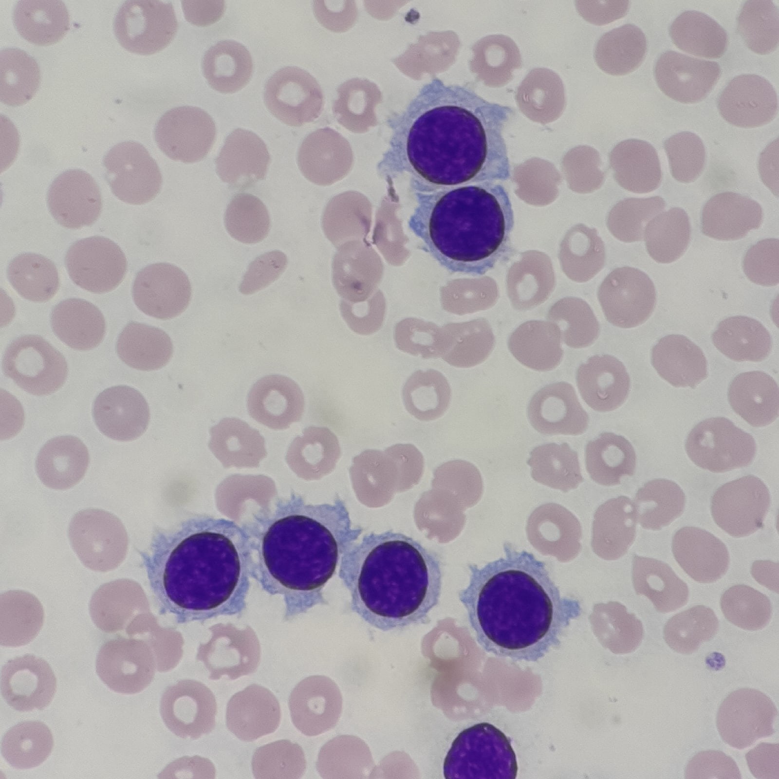
Woman, 91 years old, analysis from a polyclinic HGB - 129 g/L RBC - 4.44 million/µL PLT - 105 thousand/µL WBC - 99.6 thousand/µL 84% hairy cells (we don’t perform immunophenotyping, but I don’t think anyone will have doubts) The analyzer initially counted them as monocytes The photos show two different Romanowsky stains, manual (more pink) and automated, 100x magnification and phone zoom
Hairy Cell Leukemia: Blood Picture and Cell Morphology
A clinical case of hairy cell leukemia: leukocytosis up to 99.6×10⁹/L, morphology of hairy cells in a blood smear, and analyzer scattergram.