

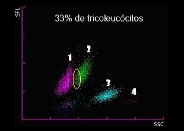
When performing flow cytometry (leukocyte distribution diagram), in which cell area are tricholeukocytes identified?
1 — lymphocytes 2 — monocytes 3 — neutrophils 4 — eosinophils
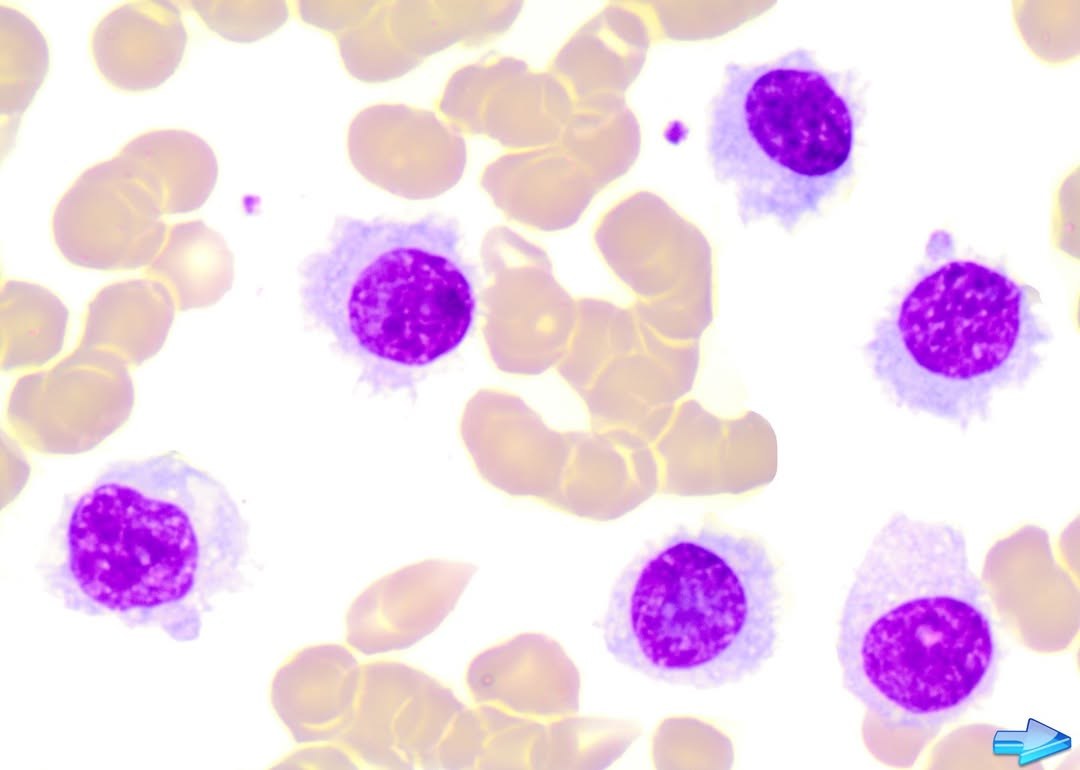
Figure 1. Variant HCL: 9000 leukocytes per cubic millimeter, 33% hairy lymphocytes. Figure 2. Leukocyte distribution diagram in this case.
🔬 Source: from the archive of Brazilian hematology specialist Marcio Melo


