

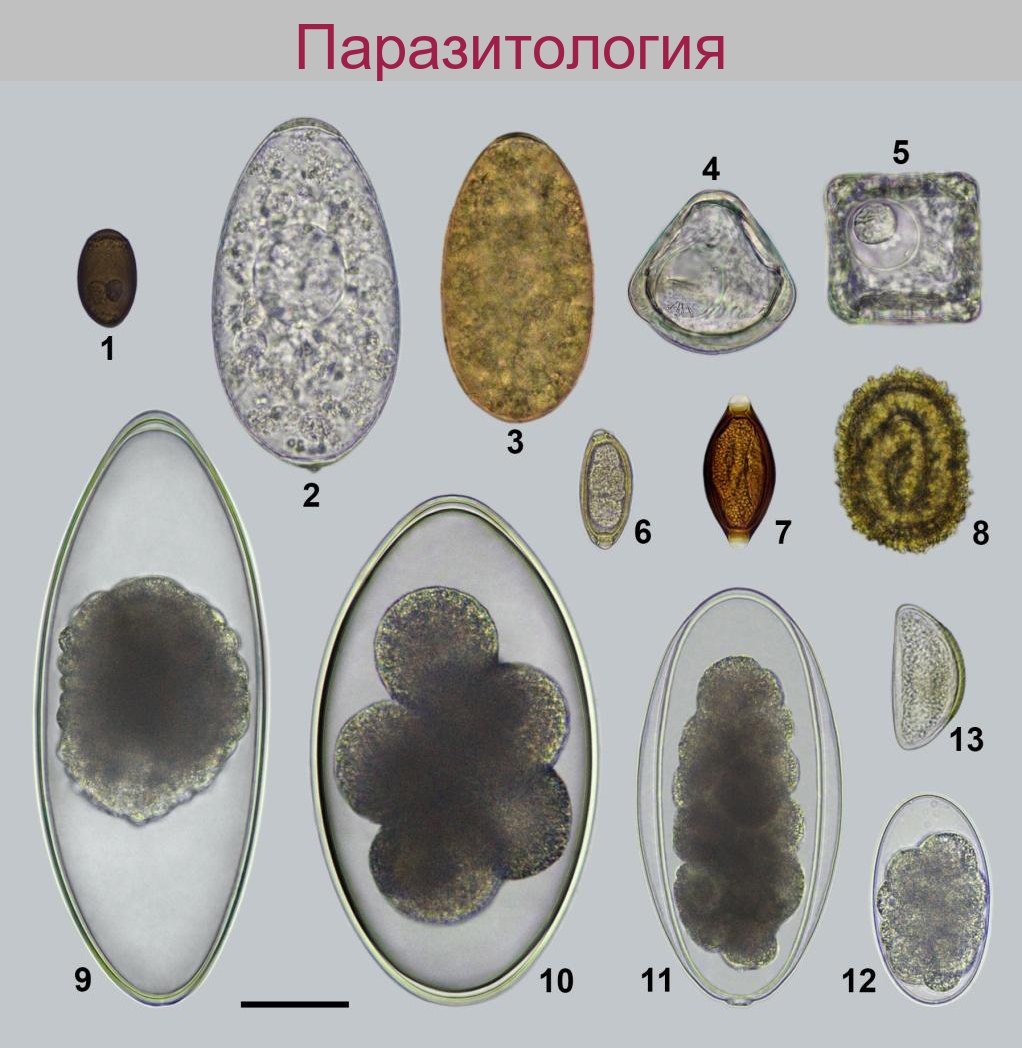
Helminth eggs detected in reindeer by coproscopy: 1 – trematode egg of the genus Dicrocoelium; 2 – trematode egg of the genus Paramphistomum; 3 – trematode egg of the species Fasciola hepatica; 4 – cestode egg of the species Moniezia expansa; 5 – cestode egg of the genus Moniezia; 6 – nematode egg of the genus Capillaria; 7 – nematode egg of the genus Trichuris; 8 – nematode egg of the species Ascaris mosgovoyi; 9 – nematode egg of the species Nematodirella longissimespiculata; 10 – nematode egg of the genus Nematodirus; 11 – nematode egg of the genus Marshallagia; 12 – nematode egg of the order Strongylida; 13 – nematode egg of the species Skrjabinema tarandi. Light microscopy, scale bar length is 0.05 mm. All eggs are presented at a uniform scale.
Source: Loginova O. A. Diagnosis of Helminthiasis in Reindeer, 2024. 💎 Find more useful materials on parasitology in our LAB-TESTS library. A link with access to the library is automatically sent to you via private message after paying for a VK donut Labformula subscription.


